

Pterygium Surgery: Purpose, Procedure, and Benefits and Side Effects
Last Updated: Feb 28, 2023
What is Pterygium surgery?
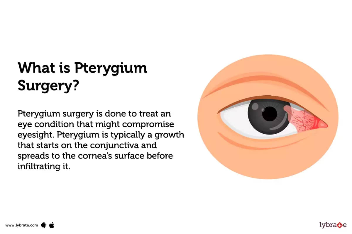
Pterygium surgery is done to treat an eye condition that might compromise eyesight. Pterygium is typically a growth that starts on the conjunctiva and spreads to the cornea's surface before infiltrating it. It usually resembles a triangle as it develops, with the pterygium's head toward the center and the body and tail (the triangle's base) toward the canthus (the point where the upper and lower eyelids meet). Usually, the increased growth starts above the cornea's edge and moves within, where it might eventually compromise eyesight.
Although the exact reason for pterygium is still unknown, studies indicate involvement of a high UV exposure and dry environment as contributing factors in the development of this condition. Patients with pterygia are commonly from warm climates or people with light color eyes and less pigmentation are also at a higher risk of developing it. Also, chances of pterygium formation can increase as a result of trauma from exposure to pollen, dust, sand, wind, and other environmental stressors.
Types of Pterygium surgery:
There are currently dozens, if not hundreds, surgical procedures that are all intended to remove pterygia and only differ in how the conjunctiva is closed after the excision. Conjunctival or conjunctival-limbal autografts are presently the 'gold standard' of pterygium surgery due to their comparatively low recurrence rates. Although the most popular procedure, simple excision, only takes a few minutes, with this method the likelihood of the pterygium returning is substantially higher. During this method, the surgeon just peels and cuts the corneal scar tissue off. It works in roughly 90% of situations, but in 10% of the cases it comes back violently.
Here are some of the many methods to remove pterygium surgically;
Excision with simple wound closure
Following the removal of the pterygium, the conjunctiva tissue transplant will either be sutured or glued in place using fibrin glue. Both methods lessen the chance of pterygia returning.
Dissolvable sutures may be a standard procedure, but they can make patients feel worse after surgery and prolong their recovery by several weeks.
On the other hand, applying fibrin glue has demonstrated to lessen pain and swelling while halving the amount of time needed for recovery (compared to using sutures). However, because fibrin glue is a blood-derived substance, there is a chance that it could spread viruses and diseases. Additionally, using fibrin glue may cost more than using sutures.
Bare sclera procedure
The bare sclera approach is an additional choice, however it entails a higher risk of pterygium recurrence. The pterygium tissue is removed by your doctor during this more conventional operation without being replaced with a tissue graft. This exposes the eye's inner white, allowing it to heal naturally.
The bare sclera approach does away with the dangers of sutures and fibrin glue, but pterygium regrowth is common and tends to grow larger.
.jpg)
Conjunctival-limbal autograft
The pathogenesis of pterygium may involve limbal stem cells in a significant way. Since pterygium frequently affects the limbal stem cells, limbal autograft may be suggested as a pterygium treatment. Conjunctival-limbal autograft is a common procedure the surgeons use to treat recurring and advanced pterygium.
Conjunctival autograft
Conjunctival autograft includes utilizing tissue to cover the area where the pterygium was removed by extracting neighboring conjunctiva and limbal tissue in one piece from another area of the patient's eye.
Conjunctival rotational autograft
In situations where it is neither feasible or acceptable to use the superior conjunctiva as a donor source, conjunctival rotation autografting is a viable approach for conjunctival grafting. It is performed following pterygium removal as an alternative to standard conjunctival autograft. In this treatment, the underlying fibrovascular pterygium tissue is removed, and the original epithelium is rotated 180 degrees over the bare sclera while still containing some subepithelial tissue.
Mitomycin C + Conjunctival or Conjunctival-limbal autograft
This antimetabolite agent helps to prevent recurrences by preventing growth at the conjunctival level. It is either applied to the bed of the sclera after the pterygium is removed, or it is given as eye drops following the procedure.
Amniotic membrane transplantation
Limbal autograft and amniotic membrane transplantation are anticipated to be successful treatments for recurrent pterygium in which partial stem cell deficiency and inflammation coexist.
Lamellar keratoplasty in conjunction with pterygium surgery
Due to its intricacy and requirement for a donor cornea, peripheral anterior lamellar keratoplasty is rarely performed. However, it is regarded as the preferred technique in individuals who face recurrences or who have strong pterygium activity. The recurrence in this method is relatively negligible.
Benefits of Pterygium surgery:
The removal of a pterygium can help prevent vision loss and additional harm to your eye from the condition. It is a fairly safe treatment because the operation only involves the outer layers of the eye. The appearance of your eye should also get better once the redness has subsided.
Here are some of the benefits of pterygium removal surgery;
- Improves eye vision
- Reduces chronic irritation and inflammation
- Treats irritation and inflammation that are not managed with eye drops
- Prevents further damage to the eye
- Prevents impact on vision due to the pterygium
- Enhance appearance.
Why is Pterygium surgery done?
Pterygium eye surgery is not required unless the growth is causing discomfort despite the use of artificial tears, is causing astigmatism or vision loss, or is getting close to the field of vision. Patients frequently choose to have the pterygium removed for cosmetic reasons. You should be warned that pterygia can sometimes grow back violently and quickly following surgery. After the removal, patients also feel dryness and irritation, although these side effects can be treated and prevented with surface lubrication and other drugs.
A pterygium can either be atrophic or progressive (degenerated). The head of a progressing pterygium is thick, fleshy, and vascular, revealing the presence of opaque infiltrates. A thin, poorly vascularized pterygium is an atrophic pterygium. The existence of the Stocker's line, a pigment line in front of the pterygium, is indicative of a long-standing, non-progressive pterygium. According to the visibility of the underlying episcleral structures, pterygium stages have been proposed.
The following situations call for pterygium surgery:
- Possibility of a progressive pterygium
- Progressive pterygium that may encroach the visual axis
- Large Pterygium causing severe astigmatism
- Recurring episodes of inflammation
- Double vision
A minimally invasive procedure takes 20 to 30 minutes to complete. The pterygium is first thoroughly peeled in order to reduce the likelihood of recurrence. After that, a tissue transplant from under your upper eyelid is removed and placed over the area that was removed. Instead of stitches, tissue glue is usually utilized to hold the graft in place.
What are the risks of Pterygium surgery?
Surgical risks and complications of pterygium surgery include:
- Recurrence of the pterygium
- Developing an infection or cyst
- Double vision that persists and needs surgery
- It's possible that your eye will remain dry or inflamed
Despite the risks, a pterygium surgery can be a good choice for people with double or compromised vision issues. The chances of countering the risks can also be ignored or reduced if the surgery is done by an expert eye surgeon at Pristyn care.
How do I prepare for Pterygium surgery?
Pterygium removal is a minimally invasive procedure and usually takes only 30 to 45 minutes. To help you get ready for pterygium surgery, your doctor may give you some general instructions, that may include;
- Before the operation, wash your hair
- On the day of the treatment, avoid applying any lotions, powders, or scents
- You might need to fast beforehand or just have a little meal before the surgery if local anesthetic is utilized
- Please inform the doctor of all medications you are taking as some, particularly blood-thinning drugs, may need to be stopped prior to the treatment
- Before the operation, use any eye drops that your doctor has advised
- Avoid wearing contact lenses for at least 24 hours before the treatment (if you wear them)
- The doctors will advise you to make arrangements for transportation following the surgery since you won't be able to drive yourself due to the light sedation
- Contact your surgeon if you have any eye allergy, infection, redness etc before the procedure. He/she might wish to see you before the surgery or postpone the appointment until after you recover.
Speak with a reputable healthcare professional of Pristyn care before the procedure to receive detailed instructions on how to get ready for the surgery. Your medical professional might be able to provide you with information about the pre- and post-surgery preparation and treatment.
How is Pterygium surgery done?
Mentioned below are the points:
Before the procedure
Pterygium removal is a minimally invasive procedure. Usually, it only takes 30 to 45 minutes. To help you get ready for your pterygium surgery, your doctor may probably give you some general instructions.
- The patient is brought to the operating room on the day of surgery well in advance of the scheduled procedure time
- Prior to the surgery all clothes and surgical instruments are cleaned to eliminate any bacteria and germs
- The patient is changed into a fresh surgical gown
- A thorough physical checkup is performed before the procedure. Vitals such as the heart rate, blood pressure, body temperature, and breathing rate are monitored
- Depending on the issue and method, pterygium surgery varies substantially. The procedure can start when you've been prepared and given the proper anesthetic
During the procedure
The pterygium can be permanently removed by excising the growth from underneath where it originates, leaving the eye looking smoother. After that, blood vessels are cauterized under a microscope.
Excision Techniques
- When removing a pterygium, a nerve blocker is frequently employed. As a result of its effective pain management during surgery, retrobulbar anesthesia is often the most comfortable for the patient
- When removing the pterygium from the surface of the eye, the surgeon takes care to avoid harming the underlying corneal tissue or removing stroma
- The limbus, where the pterygium starts to intrude over the cornea, is where an incision is first performed. By utilizing blunt dissection, the surgeon cuts it loose and peels it from the corneal surface.
- After the pterygium has been removed, the cornea is frequently polished using a diamond burr. The surgeon focuses on the sclera and conjunctiva after repairing the cornea.
To avoid recurrence, the open wound may be covered with tissue from another area of the eye using the conventional procedure of excision and grafting. If necessary, pterygium recurrence prevention measures must be added to those performed following basic surgical excision. These consist of:
- Conjunctival autograft: In this operation, a little portion of conjunctiva is extracted from either the same or the opposite eye and transplanted to the area where the pterygium was removed and a defect was left. Normal stem cells are provided by a conjunctival transplant to promote normal conjunctival development.
- Amniotic Membrane Grafting: From healthy, full-term cesarean infants, the amniotic membrane—the placenta's innermost layer—which surrounds the fetus is harvested. After being trimmed to the right size, the membrane is then spread over the area where the pterygium was eliminated. The amniotic membrane offers healthy stem cells that support healthy conjunctival development. A particular adhesive or sutures are used to affix the amniotic membrane to the uterus.
- Mitomycin C: This medication helps to prevent recurrences by preventing growth at the conjunctival level. After the pterygium is removed, it is either placed to the sclera's bed or administered as eye drops.
After the procedure
- Your medical team will transfer you to a recovery room or the post-anaesthetic care unit (PACU) so you can wake up from the anesthetic. With local anesthesia, this normally takes 10 to 15 minutes
- Your surgeon will place an eye pad or patch after the procedure to provide comfort and stop infection
- In order to minimize graft edema and increase graft survival, topical steroids are frequently utilized in the early postoperative phase
- Additionally, you can be given medications to ease your post-surgery discomfort, oral antibiotics to help prevent infection, and wound care instructions
- Your eye(s) will be covered for at least one day following the treatment. You should take a family member or friend with you to the hospital and they should also accompany you home
- The next day, you have a follow-up appointment. Depending on your doctor's recommendations, you can return to your home if you don't experience any unusual symptoms
- Please schedule routine follow-up visits after the operation. After the procedure, there should be a follow-up appointment one month later, then another one after three months, and finally one after six months.
How much does Pterygium surgery cost?
Pterygium surgery costs typically range between ₹15,000 to ₹40,000 and might vary depending on the type of hospital selected, the grade and stage of the pterygium, the recommended technique, the patient's medical condition based on age and other health considerations, etc.
What to eat after Pterygium surgery?
You must follow a high protein, vitamin rich diet following Pterygium surgery, avoiding eating food or drinks high on sugar content as they can aid in infection and slow down the healing process. So, while you're healing after Pterygium surgery, you may still indulge in a broad selection of savory and nourishing meals;
- Citrus fruits
- Red bell peppers
- Green leafy vegetables
- Milk
- Carrots
- Tomatoes
- Berries
- Broccoli
- Spinach
- Guava
- Grapefruit
- Orange
- Whole grains
- Sprouts
- Eggs
- Meat
- Fish
- No fried or junk food
- Avoid sweets.
Is Pterygium surgery safe?
The surgical technique for pterygiums is rapid and low-risk. Pterygium surgery is very safe compared to other forms of eye surgery since it only affects the outer layers of the eye.
Is Pterygium surgery painful?
Surgery for pterygiums is relatively painless than other ocular operations. Since your doctor will sedate you and numb your eyes before the process, you won't actually feel any discomfort during the procedure. However, the operated eye(s) can hurt at first, the discomfort should quickly subside during the first 24 to 48 hours following surgery. There is typically some little soreness that lasts for the next 24 to 48 hours before steadily getting better.
How long does it take to recover from Pterygium surgery?
Recovery time following pterygium removal might range from a few weeks to a few months. The technique used to do the removal and how well you take care of yourself after your treatment are two factors that impact how long your recovery will take. Keeping your eyes moist after your treatment is one of the most crucial things you must do. Use artificial tears to moisten your eyes if they feel dry.
It's crucial to shield your eyes from dry and dusty environments. You should wear adequate eye protection if you find yourself in such circumstances. Additionally, while you are recovering from your treatment, it is crucial that you shield the sun from your eyes. Wear wraparound sunglasses to totally shield your eyes from UVA and UVB rays whenever you are exposed to sun.
Your doctor will give you aftercare advice, such as how to clean properly, how to take antibiotics, and how to arrange follow-up appointments.
What are the side effects of Pterygium surgery?
There are complications and side-effects in any medical treatment. It's common to feel some pain and have some redness after the pterygium surgery. It's also typical to experience some blurriness when recovering. However, there are some other side effects related to Pterygium surgery that may include;
- Poor eyesight
- Recurrence of the pterygium
- Graft edema
- Graft loss
- Graft sliding
- Granuloma
- Corneal ulceration or infection.
Pterygium surgery Aftercare:
- It's crucial that you carefully follow your doctor's recommendations after the treatment
- After the surgery, the doctor will prescribe eye drops, use them as directed on a regular basis to avoid infection and inflammation
- For at least six weeks following the treatment, keep the afflicted eye properly lubricated and stay away from irritants as much as possible
- For two weeks following surgery, use your eye shield at night to protect yourself from injury
- Make sure to wash your hands before touching the area around your operated eye(s)
- If your eyelashes have any crusting, gently remove it by wetting a cotton ball in saline solution
- Avoid rubbing, pressing, or bumping the operated eye in order to injuring your eye and also to prevent displacing the connected tissue
- Limit your time outside and your time staring at a computer, television, book, screen or your exposure to the wind and sun. To lessen the chance of recurrence, wear sunglasses outside to shield your eyes from the sun and wind
- After surgery, refrain from using eye makeup or mascara for two weeks
- The doctor will remove the sutures that were applied about 7 to 10 days following the surgery and give instructions on how to avoid a recurrence
- You might notice that the affected eye has more blood vessels than usual in the first month following the surgery, giving the eye a rather red appearance. That is typical. It might make you feel uneasy. Make sure to use artificial tears to lubricate the eye and prevent irritation by wearing sunglasses to shield the eye from wind and light
- Your eyes may feel scratchy and irritated, and your vision may change slightly. If something doesn't seem to be healing regularly, consult your doctor right away.
Conclusion:
Pterygium may appear strange and even frightening, but it is not a very serious eye condition and doesn’t even need medical attention. If the condition starts to cause eye discomfort it can be relieved by using artificial tears, steroid eye drops, or ointment. However, your eye doctor can employ surgical intervention if the pterygium expands and impairs your vision or if you don't like the way your eye appears. When you're outside, even on cloudy days, don't forget to wear sunglasses with UV radiation protection for your eyes from developing Pterygium.
Table of content


15+ Years of Surgical Experience
All Insurances Accepted
EMI Facility Available at 0% Rate
Find Ophthalmologist near me

Ask a free question
Get FREE multiple opinions from Doctors