Breasts (Female Anatomy): Image, Function, Diseases, Treatments
Last Updated: Apr 05, 2023
Breasts Image
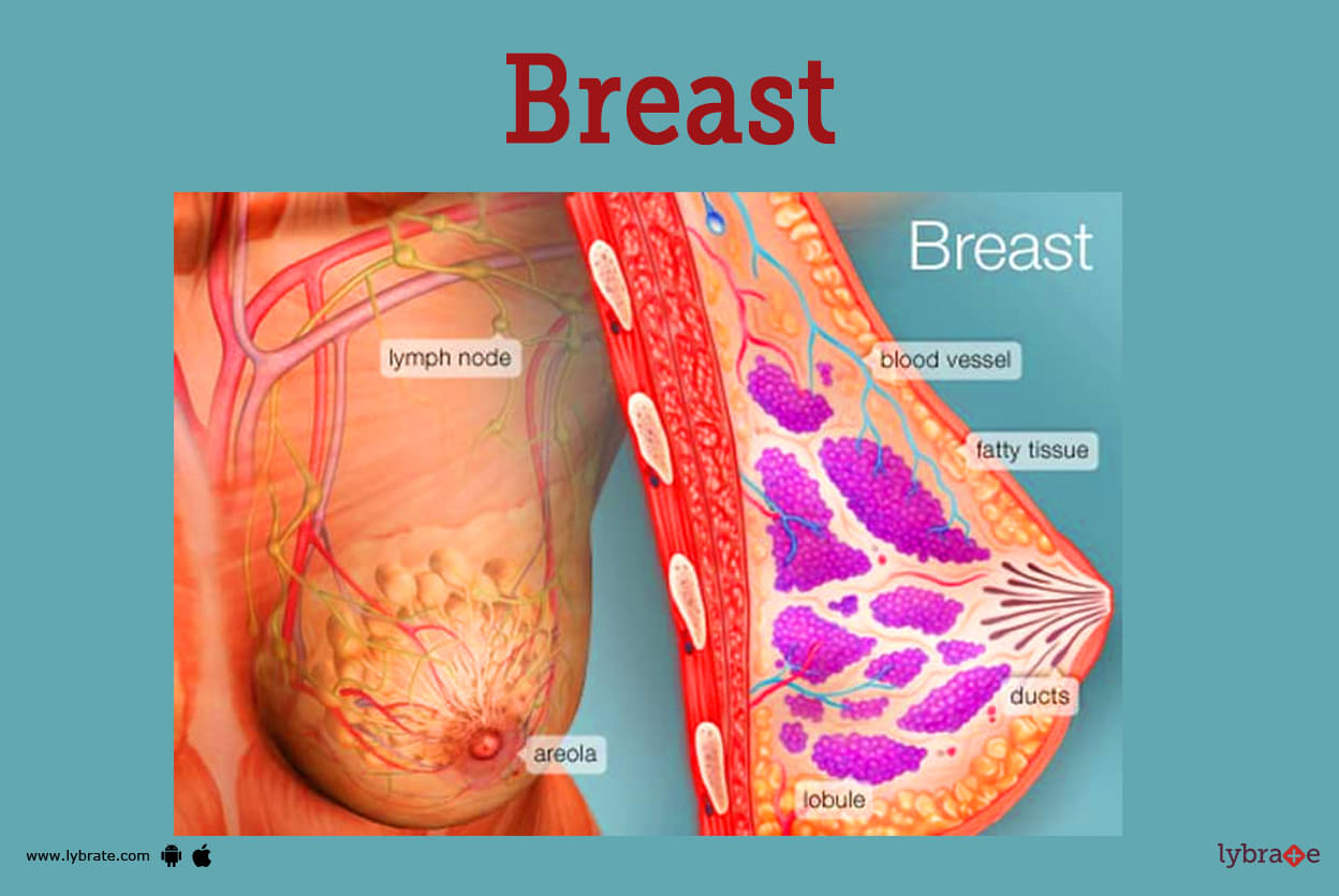
- It is a cushion of the tissue overlying the chest muscles. Breasts consist of specialised tissue that is important from the point of producing milk (glandular tissue) and also a collection of fatty tissue.
- Since every female has a different body and shapes, the size of their breasts varies from person to person. Since the uptake of food varies from person to person, also depending on the conditions in which she is living, their size may vary.
- It starts from the mammary lobes that are around 15 to 20 in sections. The part after the lobe is the mammary lobules. These lobules are the parts where milk is produced. The next part are the mammary ducts, which are collections of mammary lobules.
- The milk travels through these networks of ducts. The collection of all the mammary ducts than unites and opens out into nipples through larger ducts, which finally exit the skin in the nipple. Areola is the dark colour of skin that surrounds the nipples.
But how is this tissue so well supported in its place? Well, it is due to the presence of the connective tissue and the ligaments that provide support to these breasts and give them such a beautiful shape and size. A sensation is also present in them due to the presence of nerves.
Breasts Functions
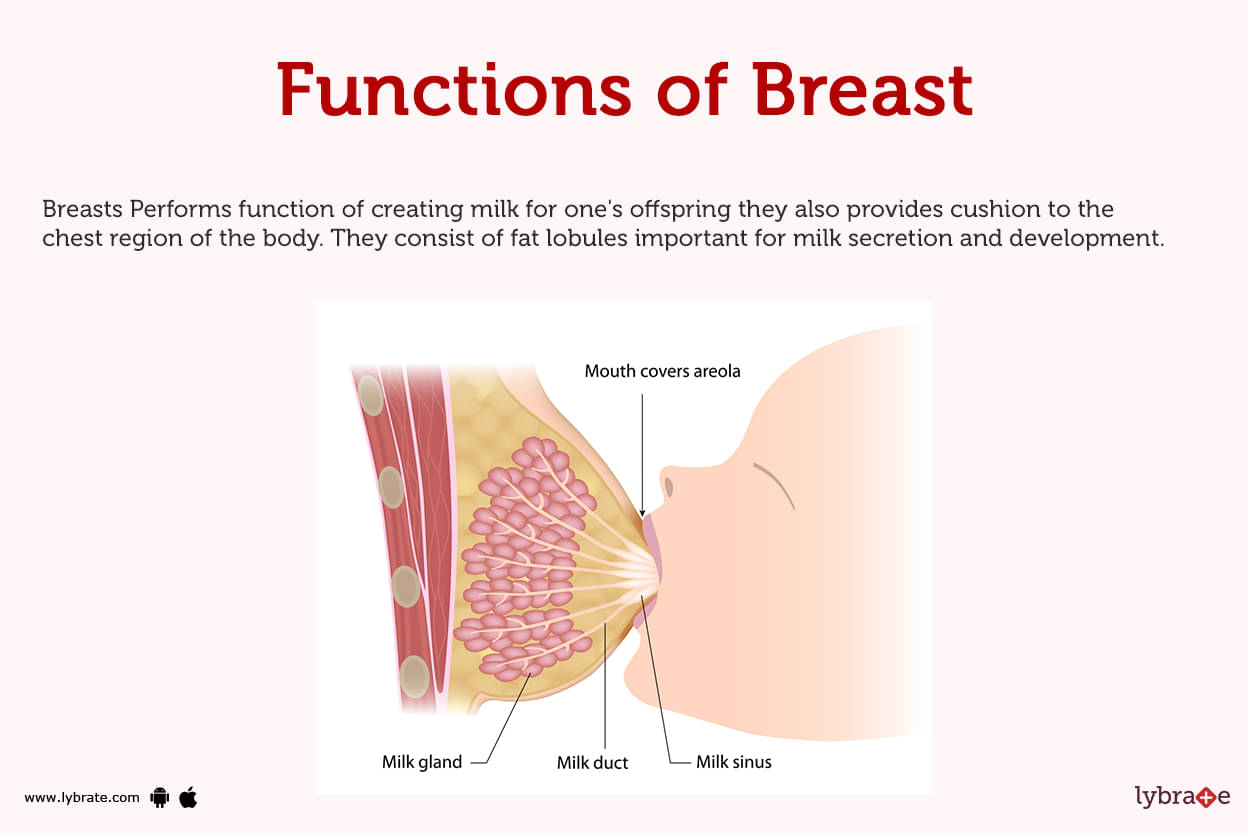
Breasts Performs function of creating milk for one's offspring they also provides cushion to the chest region of the body. They consist of fat lobules important for milk secretion and development.
During pregnancy the breasts grow further. This growth is much more uniform than that at adolescence. The breasts of women with small breasts tend to grow about as much during pregnancy as those of women with large breasts.
- Nipple: The nipple becomes erect because of such stimuli as a cold environment, breastfeeding, and sexual activity. The nipple of the post-partum female is used by the infant to breastfeed.
- Areola: The small darkened (pigmented) area around the nipple is called the areola. (The word 'areola' is the diminutive of the Latin 'area' meaning a small space.) In pregnancy the areola darkens further and spreads in size. The areola contains small modified sweat glands (Montgomery's glands) that secrete moisture that acts as a lubricant for breastfeeding.
Breast Diseases
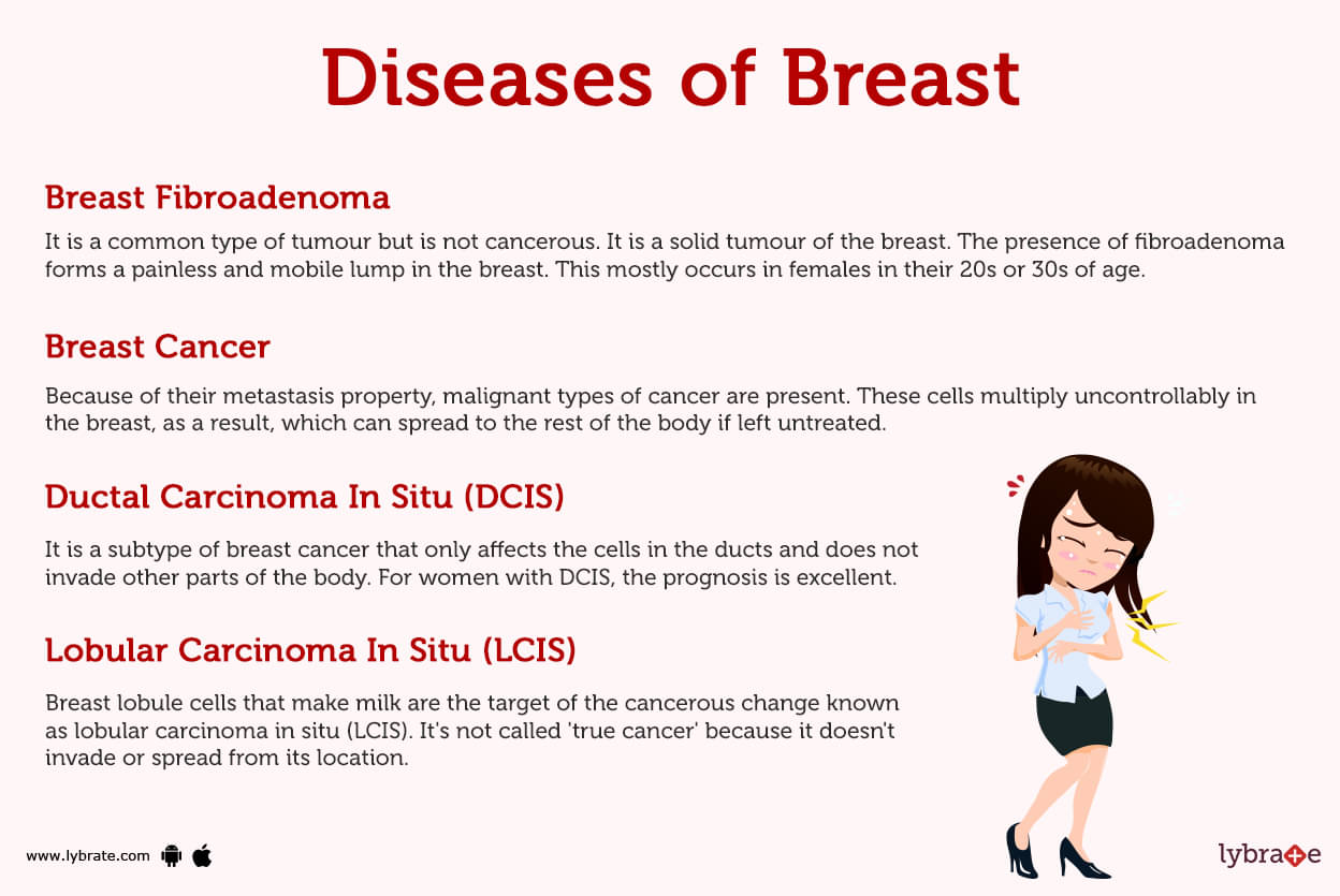
- Breast Cancer: Because of their metastasis property, malignant types of cancer are present. These cells multiply uncontrollably in the breast, as a result, which can spread to the rest of the body if left untreated. If found in the presence of breast cancer, then symptoms like lumps, bloody nipple discharge, and skin changes can be seen.
- Ductal Carcinoma In Situ: It is a subtype of breast cancer that only affects the cells in the ducts and does not invade other parts of the body. For women with DCIS, the prognosis is excellent.
- Lobular Carcinoma In Situ (LCIS): Breast lobule cells that make milk are the target of the cancerous change known as lobular carcinoma in situ (LCIS). It's not called 'true cancer' because it doesn't invade or spread from its location. Although women who have LCIS have a higher risk of acquiring invasive breast cancer in the future, LCIS itself does not cause invasive breast cancer.
- Invasive Ductal Carcinoma: This is a type of cancer that depends on the breast but is started in the duct cells and can spread to all over the body tissues, 'a property that we call metastasis'. This has been identified as the form of invasive breast cancer that occurs in the majority of patients.
- Invasive Lobular Carcinoma: It all starts in the cells of the breast lobules that are responsible for milk production. Then it goes deep into the breasts and spreads all over the body (a property we call metastasis).
- Simple Breast Cyst: This is a benign (not cancerous) form of tumour. It is characterised by a fluid-filled sac which commonly develops in females at the age of 30s or 40s. The presence of such cysts can even cause tenderness and may be drained.
- Breast Fibroadenoma: It is a common type of tumour but is not cancerous. It is a solid tumour of the breast. The presence of fibroadenoma forms a painless and mobile lump in the breast. This mostly occurs in females in their 20s or 30s of age.
- Fibrocystic Breast Disease: A specific type of tumour in which non cancerous lumps in the breasts can cause discomfort. Not only that, but it can also change in size during the menstrual cycle.
- Usual Hyperplasia Of The Breast: Biopsy of breast tissue can even show the normal appearance of cells and noncancerous ductal cells that divide abnormally. In cases of not usual hyperplasia in females, it can increase a woman's life risk of breast cancer, but the chances of such a condition are low.
- Atypical Hyperplasia Of The Breast: A breast biopsy may occasionally uncover a disease known as atypical ductal hyperplasia or atypical lobular hyperplasia in which the cells of the breast ducts or lobules divide and do not seem normal. Both of these conditions are associated with breast cancer. Such a condition is not found to be cancerous.
- Intraductal Papilloma: This is a noncancerous condition in which breasts are found with a wart-like appearance due to the growth of this mass inside the ducts of the breasts. Intraductal papillomas can be felt in the form of a lump. In certain cases, a clear or bloody fluid can even leak out from the nipple.
- Adenosis Of The Breast: This is a type of noncancerous condition due to enlargement of the lobules of the breasts. A mammogram may indicate a possibility of breast cancer; hence, a biopsy may be required to rule out the possibility of breast cancer in the event that there is any room for uncertainty.
- Phyllodes Tumour: This type of tumour is common and looks large. It is a rapidly or fast-growing breast tumour which looks like fibroadenoma on taking an ultrasound. It can either be benign or malignant. It is found in females in their 40s.
- Fat Necrosis: A lump in the form of a scar is developed when an injury is present in the fatty part of the breast.
- Mastitis: Mastitis is due to the presence of inflammation in the breast that shows symptoms like redness, pain, warmth, and further inflammation. Although nursing mothers are at a high risk for mastitis, which is the result of infection.
- Breast Calcifications: When mammograms are done, there are calcium deposits that can be seen in the breast called breast calcifications. The calcium pattern may suggest cancer that further leads to tests or a biopsy.
- Gynecomastia: This is a condition of overdevelopment of male breasts due to the presence of an extra X chromosome that leads to such an anomaly. It can affect newborns, boys, and men.
Breast Tests
- Physical Examination: Examine the breasts and nearby tissue for any kind of abnormality like lumps, skin changes (colour or touch), nipple discharge (if present) or lymph nodes. It is necessary to note the breast lumps (size, shape, and texture mainly).
- Mammogram: A mammogram is the most common test that is usually used when there is any doubt regarding the presence of a type of lump in the breast. In addition to taking low-dose X-rays, the equipment applies pressure to each breast. It is the most commonly used test because of its early detection/screening in cases of breast cancer.
- Digital Mammogram: The electronic images of both the breasts are stored in a mammogram, i.e., a computer-readable format. Standard film mammograms, on the other hand, produce pictures by creating them directly on film. This method is distinct from that.
- Diagnostic Mammogram: In order to get an accurate diagnosis of a breast anomaly or an abnormal mammogram, it may be required to do additional mammogram views on top of those that are performed during a standard mammogram.
- Breast Ultrasound: During this procedure, a device is put on the patient's skin, and high-frequency sound waves are sent through the patient's breast tissue. In the presence of medical professionals, the signals are then translated into images that are shown on a television screen in order to see the anatomical structures contained inside the body. Such a type of ultrasound can even determine whether the lump is made of fluid or solid material.
- Breast Magnetic Resonance Imaging (MRI scan): This is the most commonly used method since the detection is very accurate. A high-powered magnet and a CPU are used in an MRI scanner in order to provide detailed pictures of the breast and the tissues that surround it. Breast MRIs can supplement mammograms with new information, and they are only advised in certain instances.
- Breast Biopsy: A mammography or other imaging test is performed, and a tiny sample of tissue from the breast's abnormal-appearing region is removed and checked for cancer cells. A biopsy can be performed using a needle or with little surgery.
- Fine Needle Aspiration (Fna) Breast Biopsy: The fluid is removed from the breast tissue by inserting a small needle into the part of the breast that seems abnormal. It is the most straightforward sort of biopsy and is used for breast tumours that are easily palpable.
- Core Needle Breast Biopsy: A tube-shaped portion of breast tissue for the core is taken out after a hollow, bigger needle is placed into a breast mass.
- Stereotactic Breast Biopsy: Biopsy in which the doctor can precisely locate the suspicious breast tissue to get a sample thanks to digital images
- Surgical Biopsy: This is a very rare case in which it is recommended to take out a part or all of the breast lump to check for the presence of cancer.
- Sentinel Node Biopsy: Such a biopsy is required in the event of a primary tumour that is most likely to spread so that the medical professional may find and remove the lymph node or nodes in order to prevent the tumour from further spreading.
- Ductogram (Galactogram): Contrasting dye is injected into the breast through a duct in the nipple, and a small plastic tube is placed to allow the medical practitioner to observe the breast ducts. Finding the cause of bloody nipple discharge may be aided by a ductogram.
- Nipple smear (Nipple Discharge Exam): When abnormal fluid, such as blood mixed with milk, is released from the nipple, a sample of this fluid is taken and examined under a microscope to check for the presence of any potential cancer cells.
- Ductal Lavage: Prior to getting collected and analysed for cancer cells, sterile fluid is pumped into the nipple ducts. This investigational test is only given to women who are known to be at high risk of developing breast cancer.
.jpg)
Breast Treatments
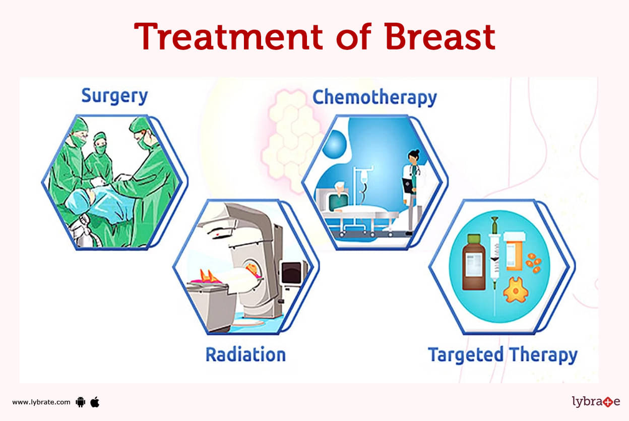
- Lumpectomy: It is a Surgery to remove a breast lump that can be a breast cancer and also some of the normal tissue that surrounds the breast. The early breast cancers are surgically removed by this and not by the method of mastectomy.
- Mastectomy: The entire breast is surgically removed. In a radical mastectomy, the surrounding lymph nodes are also removed in addition to a portion of the chest wall muscle.
- Axillary Lymph Node Dissection: When the lymph nodes of the armpits are affected by breast cancer these lymph nodes are surgically removed as these lymph nodes are the entry gate or a chance for the cancer cells to spread to the rest of the body.
- Modified Radical Mastectomy: It block removal of areolar skin and tissue and mass with the level of first second and third
- Chemotherapy: Along with the radiation, medicines are prescribed or given through the veins to kill cancer cells. Chemotherapy can be used to shrink a tumour or lessen the likelihood that it will spread or come back.
- Radiation Therapy: External beam radiation is the use of very high energy waves directed by a machine at the site of breast cancer as well as the chest wall and armpit to kill the remaining cancer cells after surgery.
- Brachytherapy: It is an additional method of radiation therapy, and it includes inserting radioactive material into a patient's body. Brachytherapy is one technique to treat cancer.
- Breast Reconstruction: A breast can be reconstructed using an implant or tissue from your own body when an entire breast or large amounts of breast tissue are removed, such as after a mastectomy.
- Breast Augmentation: Artificial implants are used during breast augmentation surgery to enlarge or reshape the breasts.
- Breast Reduction: The purpose of breast reduction surgery would be to reduce breast size. This is frequently done in women to ease neck or back pain caused by very big breasts. Men who have gynecomastia may also seek breast reduction surgery.
Breasts Medicines
- Antibiotics for Breast Infection: Since mastitis has a potential of being caused by a bacterial infection, antibiotics are often used to treat the illness.
- Calcium Medicine for Increasing Breast Size: Reduces skeletal problems and may enhance other therapies of anticancer effects. For example Bisphosphonates
- Tamoxifen for Breast Cancer: For postmenopausal women with cancers expressing oestrogen receptors whose nodes are positive or whose nodes are negative but with big tumours or poor prognostic characteristics, tamoxifen- adjuvant treatment or an aromatase inhibitor (anastrozole, letrozole, exemestane) is employed.
- Chemotherapeutic Medicines for Breast Cancer: Patients with locally advanced breast cancer benefit from neoadjuvant combination chemotherapy, which consists of the medicines cyclophosphamide, doxorubicin, and 5-fluorouracil. This is followed by surgery and breast radiation treatment.
- Combined Therapeutic Medicines: It is a medicine conjugation that shows anticancer efficacy and targets HER2-expressing cells some of the examples are Trastuzumab and emtansine.
- NSAIDs: NSAIDs, or nonsteroidal anti-inflammatory medicines, are a category of medication that is used to treat aches and pains in the knee as well as in other areas of the body. In our bodies, it can also be used as a treatment for fevers. Ibuprofen, aspirin, and naproxen sodium are examples of typical medications that fall into this category.
- Ointments and creams for Breast Pain: These creams are intended to be applied topically to the skin. This medicine provides pain relief that is effective but only lasts for a short period of time. Medicines belonging to this class that are commonly used include salicylates, counterirritants, and capsaicin.
- Corticosteroids for Breast Pain: In this treatment, an injection containing corticosteroid is injected into the knee joint. This helps to reduce the inflammation that is present in the knee joint. It can also be used to alleviate certain types of arthritis. Having said that, there are times when it is beneficial.


Table of content


Find Endocrinologist near me

Ask a free question
Get FREE multiple opinions from Doctors