Skull (Human Anatomy): Image, Function, Diseases, and Treatments
Last Updated: Feb 25, 2023
Skull Image
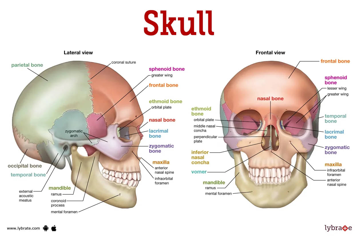
Skull, the skeletal framework of the vertebrate head made up of bones or cartilage that forms a unit that protects the brain and some sensory organs.
Skull is made up of numerous bones that are linked together by sutures and created via intramembranous ossification. Sutures are fibrous connections between the bones.
The skull bones are divided into two categories.
A baby's skull is soft, but as the bones mature, they create a thick, fibrous membrane and gradually merge to form a single skull, with the mandible being the only bone that is isolated from the rest of the skull.
- Cranium: It is also known as neurocranium. It is composed of the top of the skull and the cover, and it protects the brain, meninges, and blood vessels.
- Frontal bone: This bone runs from the forehead to the coronal suture and joins the parietal bones.
- Ethmoid bone: The ethmoid bone is located inside the eye cavity and directly beneath the nasal bridge. The ethmoid bone is a tiny, rectangular bone.
- Occipital bone: The rear of the skull is formed by the occipital bone. It joins the occipital condyles to the foramen magnum.
- Parietal bone: The parietal bone is located on the side of the skull.It is found just beneath the frontal bone, below the eye sockets and nose.
- Temporal bone: The temporal bone is responsible for the interior of the skull's sidewalls. It contains the cheekbone, ear canal, styloid and mastoid processes, and 2 temporal bone points.
Skull Functions
It is a skeletal structure that holds up the face and functions as a safe space for the brain and the eye sockets. It also protects the nerves and blood arteries that feed and innervate the brain, face muscles, and skin.
Aside from that, it permits several veins and nerves, including the cranial nerves, to enter. The foramen magnum permits the spinal cord to enter the vertebral column's spinal canal via the base of the skull.
Skull's Sutures:The coronal suture connects the frontal bone to the two parietal bones on both sides of the skull.The sagittal suture is what connects the parietal bones.The lambdoid suture serves as the connection between the occipital bone and the two parietal bones.
Fontanelles are membranous spaces between the bones that form when the sutures are not entirely fused. At the point where the coronal suture and the sagittal suture meet is where one will find the frontal fontanelle. At the point where the sagittal and lambdoid sutures meet is where one may locate the occipital fontanelle.
Skull Diseases
- Mandibular fracture: happens immediately on the side of trauma as well as indirectly on the contralateral side as a result of transferred forces. Symptoms include discomfort at the fracture site and tooth malocclusion.
- Zygomatic arch fracture: A zygomatic arch fracture is caused by trauma to the side of the face. It may cause ipsilateral paraesthesia of the face, nose, and lips by damaging the adjacent infraorbital nerve.
- Bicoronal synostosis: Bicoronal synostosis is characterised by a flattened and raised forehead in infants.
- Coronal synostosis: flattening on one side of the forehead, as well as changes in the contour of the eye socket and nose.
- Lambdoid synostosis: Lambdoid synostosis is characterised by flattening on one side of the rear of the skull. It may also cause the ear to be misplaced or the head to tilt laterally.
- Metopic synostosis: A triangle-shaped skull or a pointed forehead might result from this condition. It might also cause the eyes to seem to be closer together.
- Sagittal synostosis: is seen as a bulging out of the brow. The region surrounding the temples is also quite thin, giving the head an extended appearance.
- Cleidocranial dysplasia: Cleidocranial dysplasia is characterised by faulty tooth and bone development caused by abnormalities in particular genes influencing the cranial bones. There is a sloping forehead, extra bone inside the sutures, and an expanded skull. It is a hereditary disorder that causes cranial bone enlargement, resulting in a projecting forehead and wide-set eyes.
- Paget's disease: Paget's disease is a condition that affects the bones. Because of the unusual behaviour of osteoclast cells, new bone tissue is formed at a rapid pace, weakening the bone and increasing the risk of fracture.
- Fibrous dysplasia: Fibrous dysplasia is the presence of scar-like tissue instead of bone tissue caused by a mutation in bone-producing cells. It only affects one bone at a time, although in certain situations, more than one may be implicated.
- Osteomas: Osteomas are noncancerous bone growths in the skull.There are usually no symptoms, but if the growth presses on a nerve, it might impair the eyes and hearing, creating issues with their functioning.
- Dermoid cysts: noncancerous cysts in one or more of the three embryological layers of cells present in a growing foetus. Endoscopy may be used to surgically remove skull base dermoid cysts.
- Ossifying fibroma: Ossifying fibroma is a noncancerous lesion that mostly affects the head and neck. Cemento-ossifying fibroma is a fibrous, hard growth that often appears in the mouth or jaw. If not appropriately treated, it will continue to grow.
- Encephalocele: An encephalocele is a person born with a rare disorder that causes sections of the brain and its protective membranes to peek through the skull and is often linked with CSF leaking.
- Bruise: A laceration of the brain tissue is frequently accompanied with edoema and an increase in the pressure inside the skull, which is referred to as intracranial pressure. It raises pressure inside the skull and the brain.
- Hemorrhage: This is a brain ailment that happens when a blood artery in the brain rupture, causing bleeding into surrounding tissue, edoema, and elevated intracranial pressure.
- Hematoma: A hematoma is the creation of a blood clot. Clotting develops between the interior of the skull and the outside membrane known as the dura mater in an epidural hematoma.
- Shear damage: When the brain strikes the inside of the skull in a violent manner, this results in shear damage, which is also referred to as axonal injury. When nerve fibres that extend from the central body of a nerve cell are stretched or ripped, this might potentially cause irreparable damage to brain cells, which in turn results in troubles with the neurological system. The most common symptom is prolonged loss of consciousness.
- Stress: Stress can manifest itself physically or mentally. Anything that causes you to feel dissatisfied, furious, or nervous is a potential source. The physiological response of the body to an external threat or demand is known as stress. Small amounts of stress may be helpful, such as when it serves to warn you to impending danger or drives you to finish an essential activity in time.
- Headache: A headache of varied severity, which is frequently accompanied by nausea as well as sensitivity to light and sound. Headaches associated with migraines are often preceded by a variety of warning signs. Changes in hormone levels, particular foods and drinks, stress and exercise are all potential triggers.
Skull Tests
- CT scan: A CT scan of the head utilises x-ray equipment and computers to create multiple photos of the head and brain in order to discover bleeding, edoema, brain injury, and fractures. This is done in order to determine whether or not the patient has suffered from any of these conditions.
- Magnetic resonance imaging: During an MRI of the head, a strong magnetic field, radio frequency pulses, and a computer are utilised to produce detailed pictures of organs, soft tissues, bone, and any other internal body components.
- X-ray: An During a head X-ray, a small area of the patient's body is subjected to a low dosage of ionising radiation, and pictures of the patient's internal organs are produced. This allows for the detection and diagnosis of skull fractures.
- Physical examination: Physical examination may be used to identify sutures with ridges and facial abnormalities such as imbalanced features.
- Radioactive Test: Injecting radioactive material into the circulation, which is absorbed by the tumour if present, then utilising a special camera and a computer to generate a picture. It is used to find the bone tumour and identify any malignancy that has migrated to other organs.
- Positron Emission Tomography: A Positron Emission Tomography (PET) scan is used to identify changes in the growth of cells. It recognises tumour cells injected with radioactive glucose, allowing them to be compared to normal brain regions. It produces clinical imaging that is extremely precise of advanced tumours in both the central nervous system and the peripheral nervous system. It employs both MRI and CT cans to check for tumours at the base of the skull and rule out other issues that may not need surgery.
- Interventional Angiography: It displays key vascular structure and reduces blood loss during some operations, such as blocking off blood arteries to a tumour and providing crucial analysis to establish whether the pituitary gland is the origin of Cushing's symptoms, if they are present.
- Cranial Nerve Monitoring: This technique is used during surgery to detect and maintain vision, hearing, balance, facial expressions, speaking, and swallowing.
.jpg)
Skull Treatments
- Medical helmet: This kind of medical helmet is appropriate for babies with moderate craniosynostosis. This helmet is used to gradually reshape the head.
- Head surgery: Head surgery is performed to restructure the skull, alleviate increasing intracranial pressure, and enable the brain to grow and develop normally.
- Craniotomy: It is a type of surgery that is known to be effective when there is the existence of a brain tumour and any type of hemangioma in the brain. It is also known to be useful when a bigger tumour, hemangioma, or infarct is produced.
- Endoscopic Endonasal Surgery: Tumors situated in the middle section of the skull base may be removed using an incision at the base of the nose and endoscopic endonasal surgery. This procedure can cure conditions such as meningiomas, pituitary tumours, craniopharyngiomas, and juvenile angiofibromas.
- Eyebrow Craniotomy: To remove a skull-based tumour, a skull flap is removed from the brow, creating a tiny incision in the skull that allows access to the tumours in the front and frontal portions of the skull.
- Key hole surgery: In this procedure, incisions are made around the eye, ear, and nose in order to perform a minimally invasive surgery that removes tumours from the skull and the brain.
Skull Medicines
- Calcium supplement at the time of skull fracture: Calcium is a mineral that is required by the body. It ensures that bones get an appropriate quantity of calcium. Calcium drugs include Refirm, Calsure, Refirm, Topcal, Natcal, Calcium Sandoz, Calblend, Calcigen,Coecoral-D3, and Juscal Tablet.
- Analgesic drugs for pain at the time of small haemorrhages: It is a nonsteroidal anti-inflammatory drug (NSAID) known as etoricoxib, and it is prescribed to patients suffering from osteoarthritis, rheumatoid arthritis, and gout.
- Analgesic medicines like Narcotic: It is a narcotic pain drug that is used to treat moderate to severe pain, and oxycodone hydrochloride is the active ingredient.Tramadol is an opioid analgesic that is recommended to adults for moderate to severe pain.
- Antipyretics like Paracetamol: is a non-opiate, analgesic, and antipyretic that is used to treat headaches, pain, muscular pains, backaches, and fever, either alone or in combination with other drugs.
- Analgesics like Tapentadol: It is an analgesic that can be used to treat acute pain that ranges from mild to severe.
- Anti Seizures medicines for skull haemorrhages and infarcts: In the event of moderate to severe traumatic brain injury, if the patient develops seizures within the first week after the injury, the doctor may give the following anti-seizure drugs like sodium valproate, gabapentin, topiramate, and carbamazepine. Only if seizures occur are anti-seizure medications continued.
- Sedatives for preventing healing and bleeding of skull: Coma-inducing medicines such as propofol, pentobarbital, and thiopental are occasionally prescribed by physicians to induce transient commas in order to maintain patients in the persistent state of unconsciousness required for recovery to begin. blood vessels so tightly that they can't provide enough oxygen and nutrients to brain cells.
- Cerebral Diuretics for relieving cerebral edema: these are medications that lower the quantity of fluid in the tissues while increasing urine production. Diuretics are used to relieve pressure within the brain after a traumatic brain injury. Doctors often prescribe mannitol.
- Antidepressants for cerebral haemorrhages: Risperidone is used to treat some mental and emotional problems as well as to prevent additional damage and brain harm.
- Antivirals for treating cerebral infections of skull: They are often used in the treatment of encephalitis. Among them are Acyclovir, Ganciclovir, and others.
Frequently Asked Questions (FAQs)
What diseases affect the skull?
How do you heal a skull?
What are 4 types of skull fractures?
Can your skull be repaired?
How are skull injuries treated?
What causes skull damage?
What is the most common skull injury?
Can the skull heal itself?


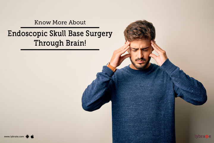

Table of content


Find Neurosurgeon near me

Ask a free question
Get FREE multiple opinions from Doctors