

Inner ear (Human Anatomy): Image, Functions, Diseases and Treatments
Last Updated: Mar 18, 2023
Inner ear Image
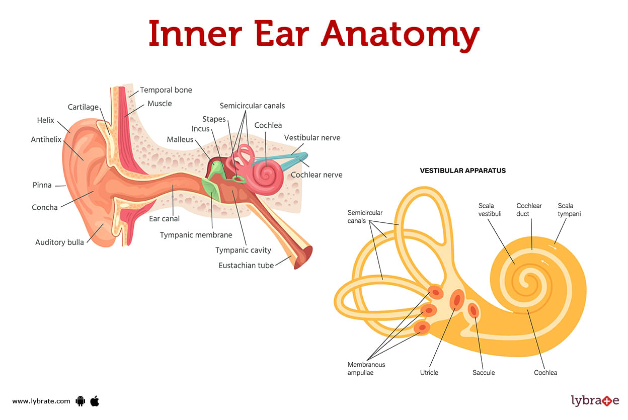
The three-part structure that makes up the inner ear is the deepest level of your auditory system. The inner ear facilitates hearing and contributes to your sense of equilibrium.
What is the inner ear?
The ear, as commonly understood, consists of three distinct anatomical elements. The visible portion of the ear and the ear canal make up the outer ear. The three smallest bones in your body are located in the middle ear, a box-shaped region behind the tympanic membrane (eardrum). The inner ear is housed in a tiny cavity in the temporal bones that help form the sides of your skull, slightly beyond the middle ear.
What are the parts of the inner ear?
The cochlea, the semicircular canals (labyrinth), and the vestibule make up the three major regions of the inner ear. The vestibule and semicircular canals help you keep your bearings, while the cochlea ensures that sound is transmitted to the brain.
What is the cochlea?
- Your fluid-filled cochlea has a snail's conical shape, narrowing from its base to its peak. Sounds with a high frequency (like birds singing) are more easily detected by the base, whereas those with a low frequency are more easily detected by the tip (like a bass drum).
- Two delicate membranes separate the cochlea into three separate tubes. The basilar membrane is one such membrane; it acts like an elastic wall and is where the organ of Corti is located.
- Tiny sensory cells called hair cells reside in the organ of Corti. The roughly 18,000 cells in your cochlea could all fit on the tip of a needle, that's how tiny they are.
- These hair cells are topped by stereocilia. Fine hair-like projections called stereocilia respond to changes in the fluid within the cochlea. Both inner and exterior hair cells exist. To better hear faint noises, the outside hair cells are more sensitive, whereas the interior hair cells are more sensitive to louder sounds.
- Although all hair cells are linked to the hearing nerve, the inner hair cell is primarily in charge of transmitting auditory information to the brain. Softer sounds are detected by the outer hair cells, which then notify the inner hair cells.
- A breakdown of the cochlea's sound-translation process is as follows:
- The small bones of the middle ear (malleus, incus, and stapes) are activated when sound enters the outer ear and strikes the eardrum (tympanic membrane) that lines the middle ear.
- Located in the cochlea's tiny oval window, the stapes are essential for hearing. The fluid in your cochlea ripples when it moves.
- The stereocilia are being swept along like seafloor plants by this wave.
- In response to this vibration, the stereocilia on the inner and outer hair cells release an electrical signal that travels via the auditory nerve to the temporal lobe of the brain. An electrical signal is interpreted by the temporal lobe as audible.
.jpg)
What are semicircular canals?
Inner ears have spiralled tubes called semicircular canals. Its canals are lined with hair cells and filled with fluid, just like the cochlea. These individual hairs are sensitive to motion rather than sound waves. The majority of non-linear motion, such as rotation, is caused by them.
What is the vestibule?
- Up and down movement is primarily controlled by the utricle and saccule in the vestibule.
- The vestibular system consists of the following parts and functions as follows:
- The fluid in your semicircular canals causes the teeny hairs in your canals to move when you move your head.
- Your saccule and utricle (the sacs that connect your vestibule to your semicircular canals) will begin to function. Your saccule and utricle, like your semicircular canals, contain fluid and microscopic hairs to detect movement.
- The vestibule and semicircular canals relay this information about motion to the brain. Then your brain relays that information to your body so that it can maintain its equilibrium.
Inner Ear Functions
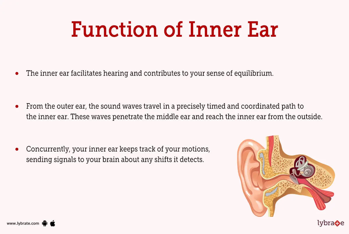
- The inner ear facilitates hearing and contributes to your sense of equilibrium.
- From the outer ear, the sound waves travel in a precisely timed and coordinated path to the inner ear. These waves penetrate the middle ear and reach the inner ear from the outside. Hearing is made possible through the transformation of sound waves into electrical energy in the inner ear, which is then sent along the auditory nerve to the brain.
- Concurrently, your inner ear keeps track of your motions, sending signals to your brain about any shifts it detects.
Inner Ear Conditions and Disorders
What inner ear conditions affect balance
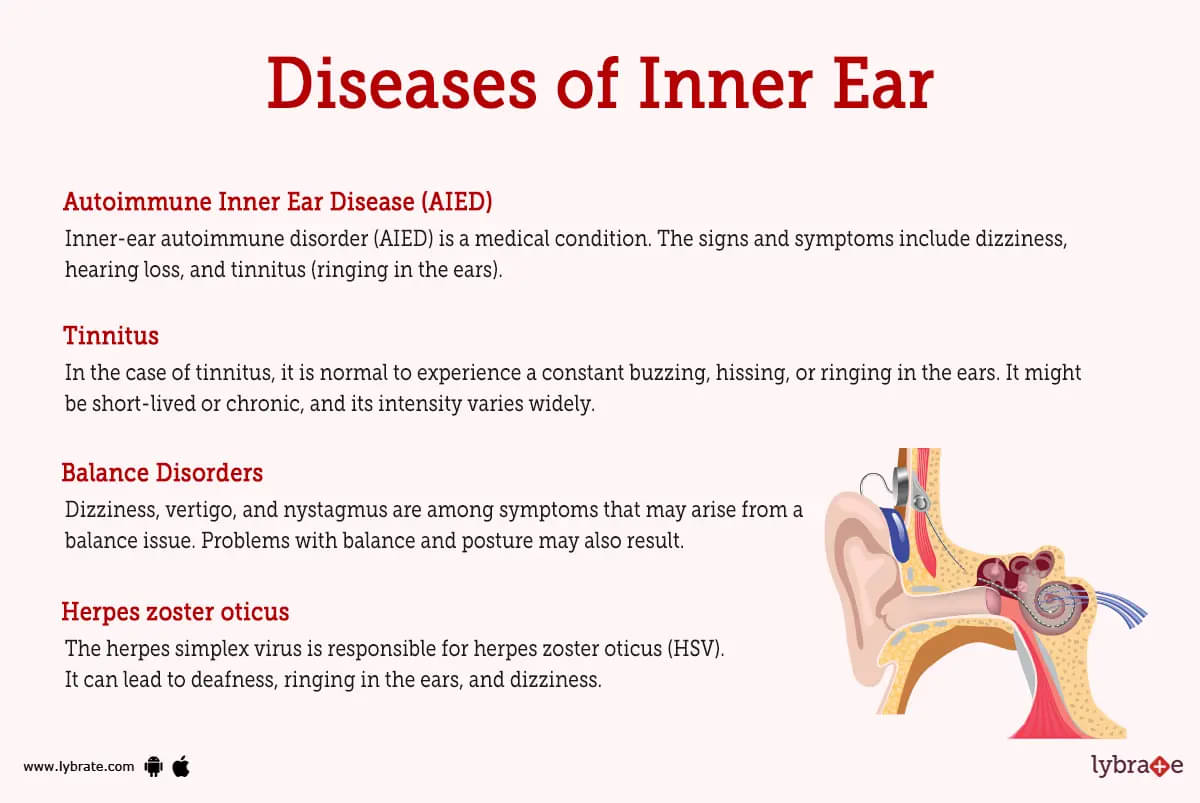
- Autoimmune Inner Ear Disease (AIED): Inner-ear autoimmune disorder (AIED) is a medical condition. The signs and symptoms include dizziness, hearing loss, and tinnitus (ringing in the ears).
- Meniere's Disease: Infrequent inner ear disorders like Meniere's disease are extremely distressing when they do manifest.
- Vestibular Disorders: Vestibular diseases are a common source of dizziness, vertigo, and nystagmus (a side-to-side or up-and-down motion of the eyes). Problems with balance and posture may also result.
- Tinnitus: In the case of tinnitus, it is normal to experience a constant buzzing, hissing, or ringing in the ears. It might be short-lived or chronic, and its intensity varies widely.
- Balance Disorders: Dizziness, vertigo, and nystagmus are among symptoms that may arise from a balance issue. Problems with balance and posture may also result.
- Benign paroxysmal positional vertigo: Disorders of equilibrium include benign paroxysmal positional vertigo (BPPV). Its side effects include lightheadedness, nausea, and throwing up.
- Drug-induced ototoxicity: Drugs like chemotherapy and ototoxic treatments can create a condition known as drug-induced ototoxicity. It can lead to deafness, ringing in the ears, and dizziness.
- Herpes zoster oticus: The herpes simplex virus is responsible for herpes zoster oticus (HSV). It can lead to deafness, ringing in the ears, and dizziness.
- Purulent labyrinthitis: Infection of the labyrinth is the root cause of purulent labyrinthitis (a part of the inner ear). It can lead to deafness, ringing in the ears, and dizziness.
- Vestibular neuronitis: Infection of the vestibular nerve leads to the illness known as vestibular neuronitis. There is a risk of deafness, tinnitus, and dizziness as a result.
- Vestibular schwannoma: Vestibular schwannoma is an infection of the Schwann cells that line the inner ear. It can lead to deafness, ringing in the ears, and dizziness.
What inner ear conditions affect hearing?
Age, disease, loud noises, heredity, and some drugs can all contribute to sensorineural hearing loss. That's the most prevalent type of hearing loss. Inaccurately called 'nerve deafness,' it is a condition that affects the ability to perceive sound. Damage to the hair cells and/or stereocilia, which carry an electrical signal representing sound to the nerve, is frequently the cause of this kind of hearing loss rather than problems with the nerve itself.
Extremely loud noises might cause permanent hearing loss. Spending time around loud noises without protecting your ears might harm or destroy your stereocilia, which detect sound waves. After being injured, hair cells and stereocilia cannot be repaired or regenerated. Hearing aids and other assistive listening devices can help those with hearing loss by amplifying sounds for the remaining hair cells and stereocilia to process.
Inner Ear Tests
Hearing test through audiogram: Audiograms are used to detect hearing loss.
- Hearing aid fitting: Once the findings of an audiogram-based hearing test have been acquired, a suitable hearing aid must be fitted to the patient. Sound will be amplified, making it easier to hear and be heard in busy places.
- Brain magnetic resonance imaging (MRI) with contrast: It is possible to see the inner ear and other brain structures with the aid of an MRI with contrast. Useful for detecting damage to the inner ear and diagnosing related diseases.
- Electrocochleography: The function of the inner ear can be evaluated with the use of electrocochleography, a test that employs mild electric shocks to stimulate the ear's auditory nerve. The vestibulocochlear nerve and hearing loss are both assessed by this test.
- Electro/Video-Nystagmography: A tiny electrode is placed on the skin over the ear, and a low-voltage electric current is passed through it to conduct the test. When the inner ear reacts, it can be captured on film. The vestibulocochlear nerve and hearing loss are both assessed by this test.
- Video Head Impulse Testing (VHIT): This procedure is used to detect vestibulocochlear nerve injury and identify hearing impairment. While your head is being moved in various directions, you will be asked to listen to a series of brief noises as part of the VHIT procedure.
- Vestibular Evoked Myogenic Potential: This procedure is used to detect vestibulocochlear nerve injury and identify hearing impairment. At certain points throughout the exam, you will be instructed to create sounds while bobbing your head from side to side and top to bottom.
- Computerised Dynamic Posturography (CDP): This procedure is used to detect vestibulocochlear nerve injury and identify hearing impairment. At various points in the CDP process, you'll be instructed to sit in a specially made chair and make a series of side-to-side, up-and-down, and forward-and-backward movements with your head.
- Audiometry (Hearing Tests): When evaluating a person's hearing abilities, audiometry is the method of choice due to the precision and accuracy it provides. It can detect hearing loss and evaluate vestibulocochlear nerve health.
- Electrocochleography (ECOG): The auditory nerve's response to sound is evaluated with the ECOG test, which consists of a series of mild electric shocks. The vestibulocochlear nerve and hearing loss are both assessed by this test.
- Computerised Axial Tomography: This procedure is used to detect vestibulocochlear nerve injury and identify hearing impairment. A sequence of digital images of the ear's inner workings are captured and analysed during this examination.
- Audiometry tests: When evaluating a person's hearing abilities, audiometry is the method of choice due to the precision and accuracy it provides. It can detect hearing loss and evaluate vestibulocochlear nerve health.
- Auditory brainstem response (ABR) testing: Auditory brainstem response testing is a mechanical method of evaluating hearing ability. It can detect hearing loss and evaluate vestibulocochlear nerve health.
- Otoacoustic emissions (OAEs): Tiny noises called OAEs occur when the inner ear is functioning normally. Both hearing loss and vestibulocochlear nerve injury can be identified and evaluated with their help.
- Vestibular test battery: The vestibular system and ocular reflexes are evaluated using a battery of tests. Videonystagmography, vestibular evoked myogenic potentials, rotary chair, and video head impulse testing are all examples of possible diagnostic procedures (vHIT). To better track eye movement, most of these exams require participants to wear goggles.
Inner Ear Treatments
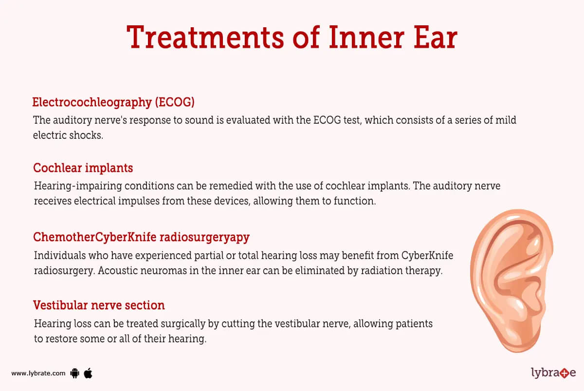
- Cochlear implants: Hearing-impairing conditions can be remedied with the use of cochlear implants. The auditory nerve receives electrical impulses from these devices, allowing them to function.
- Electrocochleography (ECOG): The auditory nerve's response to sound is evaluated with the ECOG test, which consists of a series of mild electric shocks.
- Cognitive therapy: Cognitive therapy is a treatment method that teaches patients new ways of thinking and behaving. People with hearing impairments can utilise it to enhance their ability to communicate.
- Audiologic rehabilitation: A form of therapy, audiologic rehabilitation allows those with hearing loss to recover some or all of their sense of hearing.
- Endolymphatic sac procedure: A surgical treatment called endolymphatic sac surgery can allow persons who have suffered hearing loss to recover some or all of their hearing. This process involves either destroying or draining the endolymphatic sacs in the inner ear.
- Labyrinthectomy: Labyrinthectomy is a surgical technique that can restore some or complete hearing in persons who have suffered from it. Surgery to remove the auditory cortex of the brain.
- Vestibular nerve section: Hearing loss can be treated surgically by cutting the vestibular nerve, allowing patients to restore some or all of their hearing. One's vestibular nerve in the ear must be severed.
- Acoustic Neuroma Surgery Trans labyrinthine: Surgical Treatment for Acoustic Neuroma Trans labyrinthine is a surgical treatment for restoring hearing in the deaf. The acoustic neuromas of the inner ear are surgically removed or damaged.
- CyberKnife radiosurgery: Individuals who have experienced partial or total hearing loss may benefit from CyberKnife radiosurgery. Acoustic neuromas in the inner ear can be eliminated by radiation therapy.
- Positional exercises for BPPV: Benign paroxysmal positional vertigo (BPPV) can be treated using positional exercises that aim to alleviate symptoms and restore equilibrium.
What are ways to protect my inner ears?
- It is possible to prevent damage to the inner ear and preserve your hearing in a number of ways. We'll list a few examples here:
- The volume should be lowered when watching television or listening to music, and especially when using headphones.
- If you're in an environment where loud noises are unavoidable, make sure to protect your ears with earplugs or earmuffs.
- Stay away from any events or locations that might be too boisterous. Keep your distance from any really loud sources of noise, such as speakers.
- Get away from the ear-splitting din for a while.
Inner Ear function Medicines
Steroids for reducing inflammation of Inner ear: The inflammation of the inner ear can be reduced with the use of steroids. As a result, those who are hard of hearing may be able to lead better, more fulfilling lives. Methylprednisolone and prednisone are two such instances.
- Analgesics for pain in Inner ear: Analgesics are useful for alleviating inner ear pain. As a result, those who are hard of hearing may be able to lead better, more fulfilling lives. Ibuprofen and acetaminophen are two examples.
- Muscle relaxants for stiffness in Inner ear function: Relaxants can ease stiffness in the inner ear. As a result, those who are hard of hearing may be able to lead better, more fulfilling lives. Diazepam and lorazepam are just two examples.
- Antibiotics for infection in Inner ear: Infections in the inner ear are treatable with antibiotics. As a result, those who are hard of hearing may be able to lead better, more fulfilling lives. Antibiotics like amoxicillin and erythromycin are two such instances.
- Nutritional supplements for reducing pain in Inner ear: Inner ear pain may be alleviated by taking nutritional supplements. As a result, those who are hard of hearing may be able to lead better, more fulfilling lives. Omega-3 fatty acids and magnesium are two examples.
- Supplements for promotion of growth at the time of loss of Inner ear function: Hearing-impaired individuals who take growth-promoting supplements around the time of a fracture report significant improvements in their ability to perform daily tasks and to enjoy life. Vitamin D, calcium, and zinc are just a few examples.
- Antivirals for treating infection of Inner ear: Using antivirals to treat an inner ear infection is a viable option. As a result, those who are hard of hearing may be able to lead better, more fulfilling lives. In this category, you can find drugs like acyclovir and valacyclovir.
Table of content


Find ENT Specialist near me

Ask a free question
Get FREE multiple opinions from Doctors