Get the App
For Doctors
Login/Sign-up

Health Feed
Find Doctors
Health Packages

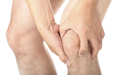
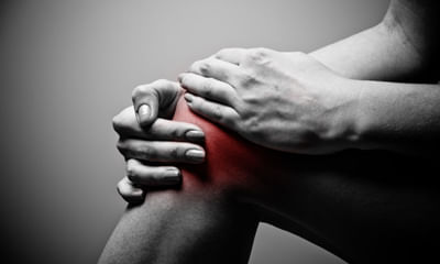
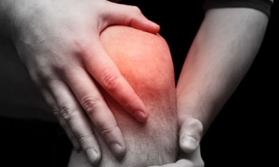
Patellar Health Feed
Health Query
Share
Bookmark
Report

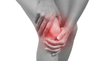
Respected Lybrate user. From the above mentioned details (mri). Very little problem is present in your both knees but don't be neglectd. Because in near future the all above's changes are converted into your knees arthritis. Please be careful. Take treatment for your orthopaedic doctor and take advices, precautions and preventive measures. In my side you are taking precautions, preventive measures, life style modifications, dietitian/nutritionist advices. Take diet chart. Ergonomical, postural c...more
59 people found this helpful
Asked for female, 43 years old from Daman
Share
Bookmark
Report

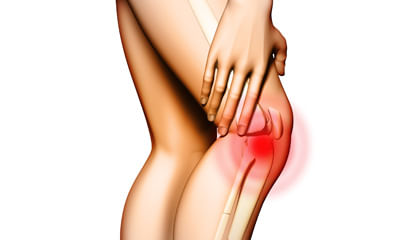
its more likely due to arthritis in the patellar cartilage. weight reduction and quadriceps strengthening exercises will help
113 people found this helpful
Asked for male, 49 years old from Kolkata
Share
Bookmark
Report

Dear friend
youmay be having some problem in your either patellar tendon, patella or the knee please get an xray of the knee done and upload for us to see and help.
youmay be having some problem in your either patellar tendon, patella or the knee please get an xray of the knee done and upload for us to see and help.
Asked for male, 19 years old from Alwar
Share
Bookmark
Report

Patellar tendon support use karo. Reduce your running for few days. If pain continues get yourself investigated with x rays and if required mri.
179 people found this helpful
Asked for male, 33 years old from Hyderabad
Share
Bookmark
Report

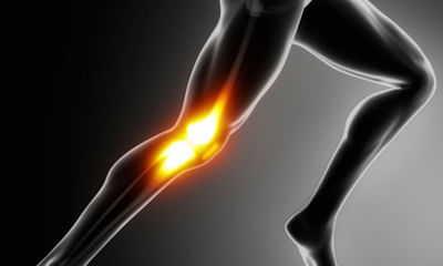
Mostly this is because of over exercising. It can also be due to certain factors like patellar malalignment, poor biomechanics etc. Good physiotherapy can help you a lot.
13 people found this helpful
Asked for Male, 35 years old from Faridabad
Share
Bookmark
Report

If the pain is at the top of bottom edge of patellar bone, it could be bcoz of hard clutch. Pain anywhere else could be something else.
132 people found this helpful
Asked for male, 16 years old from Imphal
Share
Bookmark
Report

Risk factor
a combination of factors may contribute to the development of patellar tendinitis, including:
physical activity.
Running and jumping are most commonly associated with patellar tendinitis. Sudden increases in how hard or how often you engage in the activity also add stress to the tendon, as can changing your running shoes.
Tight leg muscles. Tight thigh muscles (quadriceps) and hamstrings, which run up the back of your thighs, can increase strain on your patellar tendo...more
Asked for male, 28 years old from Mumbai
Share
Bookmark
Report
weight reduction, avoiding sitting on the floor and squatting, ice pack and quadriceps strengthening exercises help. You can also try knee brace with a patellar cut out.
156 people found this helpful
Asked for male, 24 years old from Delhi
Share
Bookmark
Report

May be a case of normal strainful patellar tendinitis.
Take rest for few days. Take warm compression and use knee cap during walking. After that if pain persist, go through a XRAY and take orthopedic consultation.
Take rest for few days. Take warm compression and use knee cap during walking. After that if pain persist, go through a XRAY and take orthopedic consultation.
55 people found this helpful
Health Query
Share
Bookmark
Report

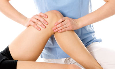
Hi sir, The pain in knee may be due to patellar tendinitis, so pain will be increasing while walking. You consult physiotherapist for treatment with ultra sound.
69 people found this helpful

Book appointment with top doctors for Patellar treatment
View fees, clinic timings and reviews

Ask a free question
Get FREE multiple opinions from Doctors
posted anonymously