Lybrate Logo
Get the App
For Doctors
Login/Sign-up
Book Appointment
Treatment
Ask a Question
Plan my Surgery
Health Feed
tab_logos
About
tab_logos
Health Feed
tab_logos
Find Doctors
tab_logos
Health Packages
Facebook Icon
Twitter Icon
Telegram Icon
Linkedin Icon

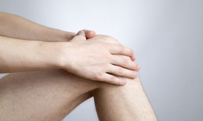
Patellar Tendonitis Health Feed

Health Query
Share
Bookmark
Report

I'm uma, 25 age. I diagnosed with pcl buckling. Please do suggest me what should I do to avoid surgery. Technique sequences pdfs, sag, coronal t2 axials findings ⚫ medial meniscus - there is thinning of medial meniscus with grade ii tear its posterior horn. • lateral meniscus grade ii tear of posterior horn of lateral meniscus. • anterior cruciate ligament - there is complete tear of acl with clumping fibres in intercondylar fossa. • posterior cruciate ligament - buckling of pcl. ⚫ medial collateral ligament - grade ii sprain of mcl. • posture-lateral corner grade I sprain of arcuate ligament. Popliteal muscle - normal -popliteo-fibular ligament - normal -popliteo-fibular ligament complex - normal short & long head of tendons of biceps femoris- normal - menisci-popliteal fascicles - normal, - lateral collateral ligament - grade ii sprain of lcl. • quadriceps tendon and patellar ligament - normal. • patella and patellar cartilage - normal. There is medial tibial-femoral joint space reduction with thinning of cartila muscles around knee - normal. Loose bodies-absent ⚫ contusions noted in posterior cortex of tibia. Mild joint effusion & suprapatellar effusion. Impression: • chronic complete tear of anterior cruciate ligament with clumping of fibres in intercondylar fossa. • grade ii tear of posterior horn of lateral meniscus. Thinning of medial meniscus with grade ii tear of its posterior horn. • grade ii sprain of medial collateral ligament. posture-lateral corner injuries as described. Mild joint effusion & suprapatellar effusion. Suggested clinical correlation and further evaluation.

Profile Image

Erasmus Mundus Master in Adapted Physica...read more

Physiotherapist•Chennai
banner-image
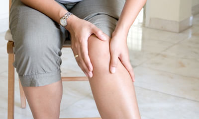
The typical symptoms of a posterior cruciate ligament injury are:
•pain with swelling that occurs steadily and quickly after the injury.
•swelling that makes the knee stiff and may cause a limp.
•difficulty walking.
•the knee feels unstable, like it may "give out"
the posterior cruciate ligament (pcl) is the strongest ligament in the knee. It extends from the top-rear surface of the tibia (bone between the knee and ankle) to the bottom-front surface of the femur (bone that ex...more
50 people found this helpful
Health Query
Share
Bookmark
Report
Profile Image

MPT - Orthopedic Physiotherapy, BPTh/BPT...read more

Physiotherapist•Noida
banner-image
Tendonitis possible and frozen shoulder possible. Meet physiotherapists for proper diagnosis and treatment.
Asked for male, 49 years old from Ambala
Share
Bookmark
Report

bachelors in physiotherapy, p.g.d.b.a (h...read more

Physiotherapist•Noida
banner-image
There are many reasons for the same can be due to knee problem, arthritis,improper footwear, injury.
Health Query
Share
Bookmark
Report
Asked for male, 23 years old from Delhi
Share
Bookmark
Report

Mrileft knee. Joint study protocol: sagittal: pd, gre// coronal: stir, tiw// axial :t2 tse findings there is mild effusion in knee joint extending into retro-patellar and lateral paracondylar recesses. Altered linear intrasubstance signal intensity is seen in posterior horn of medial meniscus appearing hyperintense on t2wand pd fs images with no intra-articular communication suggestive of grade-2 medical injury. Rests of medial and lateral meniscus appear normal. No evidence of tear. Anterior and posterior cruciate ligaments are normal in course and reveal normal parenchymal signal intensity. Quadriceps femoris tendon and patellar ligament are normal with normal signal intensity. Visualized bones reveal normal intra-medullary signal intensity and normal cortical margins. A small bony island is seen in posterior aspect of medial femoral condyle and distal femoral metaphysis. Patella is normal in location. Medial and lateral collateral ligaments are normal with no evidence of tear. Peri-articular and para-articular muscles reveal normal parenchymal signal intensity and well-preserved intermuscular fat planes. Impression: Mr. left knee finding reveals mild joint effusion with grade-2 medical injury involving posterior horn of medial meniscus as described above. Advice - please correlate clinically and with other relevant investigations please tell me 1- medicine to heal injury 2- nutrition to follow 3- physical activity during injury (i’m a gym person so workout on daily basis) 4- healing time.

Profile Image

MSPT (Master of Physical Therapy), Bache...read more

Physiotherapist•Gorakhpur
banner-image
Send your mri photo.
What problm you r facing.
Can take tab tendomac 1bd for 6 months.
Counslt to physiotherpist.
Do exercises advised by physio.
Apply ice pack n use knee brace.
44 people found this helpful
Asked for male, 23 years old from Hyderabad
Share
Bookmark
Report
Profile Image

MBBS, Basic Life Support (B.L.S), Advanc...read more

General Physician•Delhi
banner-image
The knee-jerk reflex, also known as the patellar reflex, is a simple reflex that causes the contraction of the quadriceps muscle when the patellar tendon is stretched. Let's have a detailed discussion for better advice and medication plan.
30 people found this helpful
Health Query
Share
Bookmark
Report

I have problem with my knees. I am in pain when walking, climbing stairs and exercising. I have done mri to both knees and the radiology report is as following, I need to consult an ortho doctor to discuss the mri both knees findings and have a solution: mri right knee (1.5t mri) history: pain in both knees (left > right) findings: there is minimal right knee joint effusion. Small area showing mild subchondral degenerative change in the articular part of the medial condyle of femur. Grade I changes in the posterior horn of the medial meniscus. The lateral meniscus is normal. Medial and lateral collateral ligaments are normal. Anterior and posterior cruciate ligaments are normal. Tibial, femoral condyle and upper end of fibula are normal. No evidence of loose bodies. posture-lateral corner structures including the popliteus tendon popliteo-fibular ligament are normal. Normal patella alignment is seen. The extensor tendons including the quadriceps and patellar tendons are normal. Proximal tibia-fibular joint is normal muscles surrounding the knee joint are normal. Impression: v there is minimal right knee joint effusion. V small area showing mild subchondral degenerative change in the articular part of the medial condyle of femur. V grade I changes in the posterior horn of the medial meniscus. The findings are of very early degenerative changes and are not a serious problem. Suggested clinical correlation mri left knee (1.5t mri) history: pain in both knees (left > right) there is minimal left knee joint effusion. There are few small subchondral cysts measuring 2 mm in the anterior part of the lateral tibial plateau area. There is mild suspicious focal hyperintensity in the upper part of the posterior cruciate ligament. The anterior cruciate ligament is normal. Medial and lateral meniscus is normal. Medial and lateral collateral ligaments are normal. Tibial, femoral condyle and upper end of fibula are normal. No evidence of loose bodies. posture-lateral corner structures including the popliteus tendon popliteo-fibular ligament are normal. Normal patella alignment is seen. The extensor tendons including the quadriceps and patellar tendon s are normal. Proximal tibia-fibular joint is normal muscles surrounding the knee joint are normal. Impression: v there is minimal left knee joint effusion. V there are few small subchondral cysts measuring 2 mm in the anterior part of the lateral tibial plateau area. V there is mild suspicious focal hyperintensity in the upper part of the posterior cruciate ligament. The findings are of early degenerative changes and are not a serious problem. Suggested clinical correlation there are very early degenerative changes in both knees (left > right). However, the findings are not likely to cause severe knee pain. Regards and thanks raha.

Profile Image

B.P.T, M.P.T(Orthopedics)

Physiotherapist•
banner-image
Respected Lybrate user. From the above mentioned details (mri). Very little problem is present in your both knees but don't be neglectd. Because in near future the all above's changes are converted into your knees arthritis. Please be careful. Take treatment for your orthopaedic doctor and take advices, precautions and preventive measures. In my side you are taking precautions, preventive measures, life style modifications, dietitian/nutritionist advices. Take diet chart. Ergonomical, postural c...more
59 people found this helpful
Asked for male, 19 years old from Hyderabad
Share
Bookmark
Report
Profile Image

MPT - Orthopedic Physiotherapy, BPTh/BPT...read more

Physiotherapist•Noida
banner-image
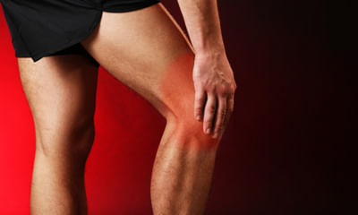
Tendonitis possible apply ice pack and pain relief ointment if it work then ok otherwise tk tens and ultrasonic therapy for 7 days.
103 people found this helpful
Asked for male, 79 years old from Pune
Share
Bookmark
Report
Profile Image

CCP, MBA, Bachelor of Ayurveda, Medicine...read more

Ayurveda•Karnal
banner-image
U need to undergo abhyanga/ swedana & patra pottali swedana. Please consult a panchakarma centre near u.
book_appt_icon
Book appointment with top doctors for Patellar Tendonitis treatment
View fees, clinic timings and reviews
chat_icon

Ask a free question

Get FREE multiple opinions from Doctors

posted anonymously
Pristyn Care Banner