Get the App
For Doctors
Login/Sign-up

About
Health Feed
Find Doctors
Health Packages

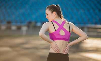
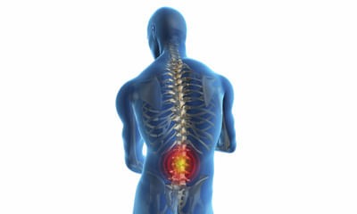
Lower back Image Health Feed
Last Updated: 6 years ago• Featured Tip
Share
Bookmark
Report

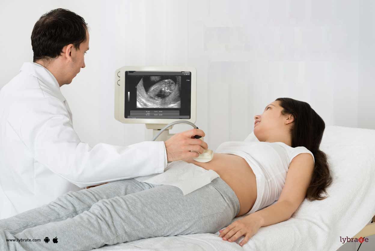
Ultra sound is commonly known as sonography. It is a process of reproducing ultrasound images of soft tissues of a particular body part and other organs on the computer screen with the help of the echoes of the sound waves produced by the transducer, a high-frequency generating instrument.
Ultra sound is commonly used during the different stages of pregnancy to denote the foetal health, date of delivery, birth defects etc. However, in recent times, the ultra sound has also been associat...more
Ultra sound is commonly used during the different stages of pregnancy to denote the foetal health, date of delivery, birth defects etc. However, in recent times, the ultra sound has also been associat...more
Asked for male, 26 years old from Patna
Share
Bookmark
Report


Common causes for changes in skin color are illness, injury, and inflammatory problems. Discolored skin patches also commonly develop in a certain part of the body due to a difference in melanin levels. Melanin is the substance that provides color to the skin and protects it from the sun. Let's have a detailed discussion for better advice and medication plan.
5 people found this helpful
Last Updated: 9 years ago• Featured Tip
Share
Bookmark
Report

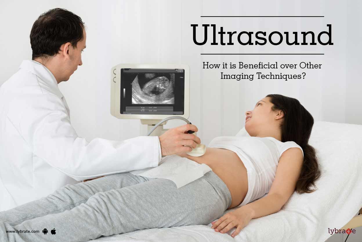
Ultra sound is commonly known as sonography. It is a process of reproducing ultrasound images of soft tissues of a particular body part and other organs on the computer screen with the help of the echoes of the sound waves produced by the transducer, a high-frequency generating instrument.
Ultra sound is commonly used during the different stages of pregnancy to denote the foetal health, the foetal sex, date of delivery, birth defects etc. However, in recent times, the ultra sound has also be...more
Ultra sound is commonly used during the different stages of pregnancy to denote the foetal health, the foetal sex, date of delivery, birth defects etc. However, in recent times, the ultra sound has also be...more
Asked for male, 46 years old from Kolkata
Share
Bookmark
Report

1.lie on your back with your legs extended and your feet flexed upward.
2.bend your right leg and clasp your hands around the knee.
3.gently pull your right leg ac
4.ross your body toward your left shoulder. Hold it there for 30 seconds. Remember to pull your knee only as far as it will comfortably go. You should feel a relieving stretch in your muscle, not pain.
5.push your knee so your leg returns to its starting position.
6.repeat for a total of 3 reps, and then switch leg...more
13 people found this helpful
Asked for female, 33 years old from Gandhidham
Share
Bookmark
Report
Hello dear there is no permanent treatment for it but injection or surgery gave long time effect in this. If you have sevear pain then go for these. If it is not too sevear then you have to consult with a good physiotherapists and take treatment of lumbar traction and ift or swd if give good relief. Even you go for blood test of vitamin b12 and vitamin d3 if there is deficiency then take supplement for it. Do regular exercise programme suggested by your physiotherapy doctor. It will subside for ...more
Asked for male, 38 years old from Visakhapatnam
Share
Bookmark
Report

Apply Hot Fomentation twice daily.
Avoid bending in front.
Postural Correction- Sit Tall, Walk Tall.
Extension Exercises x 15 times x twice daily - lying on tummy, take left arm up for 3 seconds, then bring it down, right arm up for 3 seconds, bring down. Bring right leg up, hold for 3 seconds, bring it down. Then right leg up and hold for 3 seconds and bring it down. Repeat twice a day- 10 times. Bhujang Asana -- Lie flat on your stomach, keeping the palms out, bend the neck backwar...more
Health Query
Share
Bookmark
Report

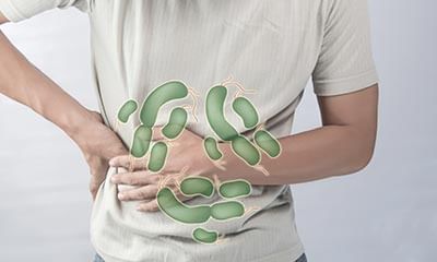
May I ask sir do yo have problem in urination.??.
Do you feel more pain after eating food ???
What is the colour of your stool ???
Since when your symptoms started.??
Sir what laxative u used.?
Try to take biscodyl 4mg/tab at before bed time for the constipation
And suggesting please drink more fluids (water or ors) if you start passing stool
And for the abdominal pain I can’t start treatment without getting my answer it can be so many cause for abdominal...more
Asked for Male, 41 years old from Hyderabad
Share
Bookmark
Report

As he had congenital, neurological deficit in the form spinal cord split there is hardly anything much to be done. He will need physiotherapy and supportive care.
Asked for male, 26 years old from Kollam
Share
Bookmark
Report
Hi, avoid high protien diet. Take tab. Drotin -m sos after meal. Do few tests ultrasound for whole abdomen. Kft, lft, urine r/m and reply soon.
43 people found this helpful
Asked for male, 29 years old from Meerut
Share
Bookmark
Report
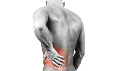
There are certain exercises like hamstring stretch, back extension and few more that gives immediate relief if done properly
consult me personally to give you images and explanation on how to go about it.
consult me personally to give you images and explanation on how to go about it.

Book appointment with top doctors for Lower back Image treatment
View fees, clinic timings and reviews

Ask a free question
Get FREE multiple opinions from Doctors
posted anonymously