Get the App
For Doctors
Login/Sign-up

About
Health Feed
Find Doctors
Health Packages

femoral artery Image Health Feed
Health Query
Share
Bookmark
Report

I am sorry to hear about your concern but will be happy to assist you.
Common causes of sudden vision loss include eye trauma, blockage of blood flow to or from the retina (retinal artery occlusion or retinal vein occlusion), and pulling of the retina away from its usual position at the back of the eye (retinal detachment).
Let's connect over a call so that we can discuss your concern in details and make a suitable treatment plan for you.
97 people found this helpful
Asked for male, 71 years old from Bangalore
Share
Bookmark
Report

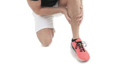
Quad Clenches: Lie flat on your back or sit upright on a chair with leg kept horizontally on another surface. Now, tighten the muscle on the front of the thigh by pushing your knee down. You should feel your thigh muscles clench, Hold for 3 secs. Repeat 10 times twice a day.
145 people found this helpful
Asked for male, 23 years old from Delhi
Share
Bookmark
Report
Send your mri photo.
What problm you r facing.
Can take tab tendomac 1bd for 6 months.
Counslt to physiotherpist.
Do exercises advised by physio.
Apply ice pack n use knee brace.
What problm you r facing.
Can take tab tendomac 1bd for 6 months.
Counslt to physiotherpist.
Do exercises advised by physio.
Apply ice pack n use knee brace.
44 people found this helpful
Last Updated: 8 years ago• Featured Tip
Share
Bookmark
Report
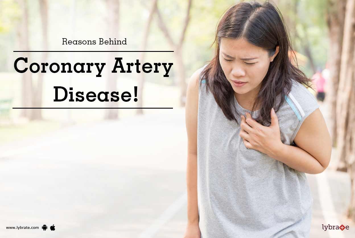
Coronary Artery Disease - Symptoms and Causes
Coronary artery disease is the disease in which the major blood vessels (coronary arteries) that supply blood, nutrients and oxygen to your heart get damaged or disease stricken. It is mostly caused due to the coronary artery getting plagued or inflamed. The consequent reduction in blood flow to the heart causes the various symptoms of coronary artery disease like angina (chest pain), short breath etc.
Symptoms:
Here is a l...more
Last Updated: 6 years ago• Featured Tip
Share
Bookmark
Report

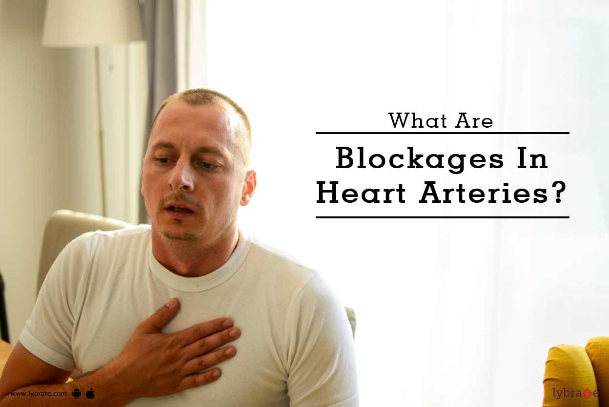
Blockage in heart is a common term used for narrowing of coronary arteries. Coronary arteries are vessels, which supply blood and thus oxygen and food) to continuously working heart muscles. Heart muscles which are not tired working from the birth till death, however, cannot sustain long without blood supply.
A reduction in blood supply gives rise to ischemia of heart muscles commonly manifested as chest discomfort or angina. A sudden complete shutdown of blood supply leads to heart att...more
A reduction in blood supply gives rise to ischemia of heart muscles commonly manifested as chest discomfort or angina. A sudden complete shutdown of blood supply leads to heart att...more
Last Updated: 7 years ago• Featured Tip
Share
Bookmark
Report

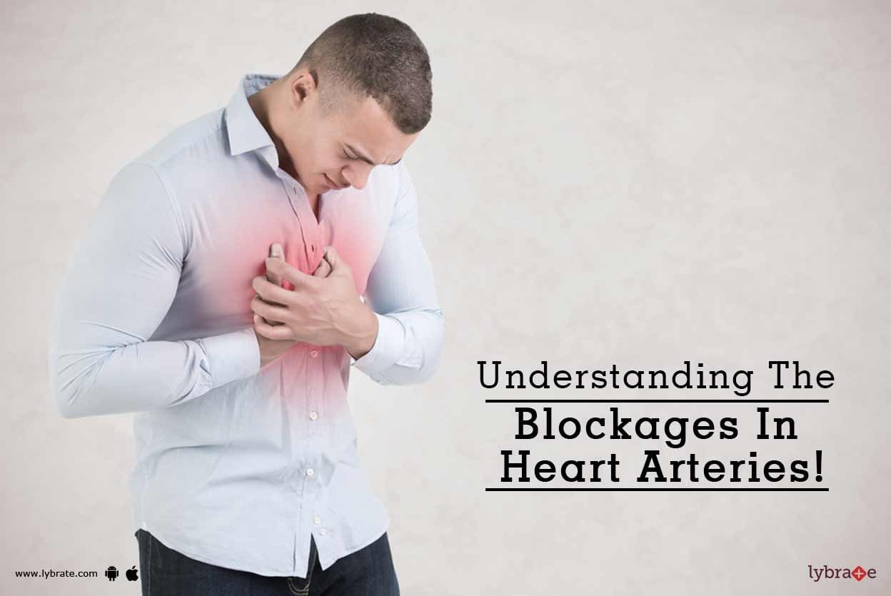
Blockage in heart is a common term used for narrowing of coronary arteries. Coronary arteries are vessels, which supply blood and thus oxygen and food to continuously working heart muscles. Heart muscles, which are not tired working from the birth till death, however, cannot sustain long without blood supply.
A reduction in blood supply gives rise to ischemia of heart muscles commonly manifested as chest discomfort or angina. A sudden complete shutdown of blood supply leads to heart att...more
A reduction in blood supply gives rise to ischemia of heart muscles commonly manifested as chest discomfort or angina. A sudden complete shutdown of blood supply leads to heart att...more
Last Updated: 6 years ago• Featured Tip
Share
Bookmark
Report

Heart Blocks are a result of plaque buildup in your arteries, which blocks blood flow and circulation to the heart, causing heart muscle damage and heightens the risk for heart attack and stroke.
Arteries which have smooth and elastic walls become thick and restrict blood flow from the cholesterol deposits over the years. Blood clots can also block the arteries that supply oxygen rich blood to the heart. These can eventually lead to strokes and heart attacks.
Some warning signs that...more
Arteries which have smooth and elastic walls become thick and restrict blood flow from the cholesterol deposits over the years. Blood clots can also block the arteries that supply oxygen rich blood to the heart. These can eventually lead to strokes and heart attacks.
Some warning signs that...more
Last Updated: 10 years ago• Featured Tip
Share
Bookmark
Report

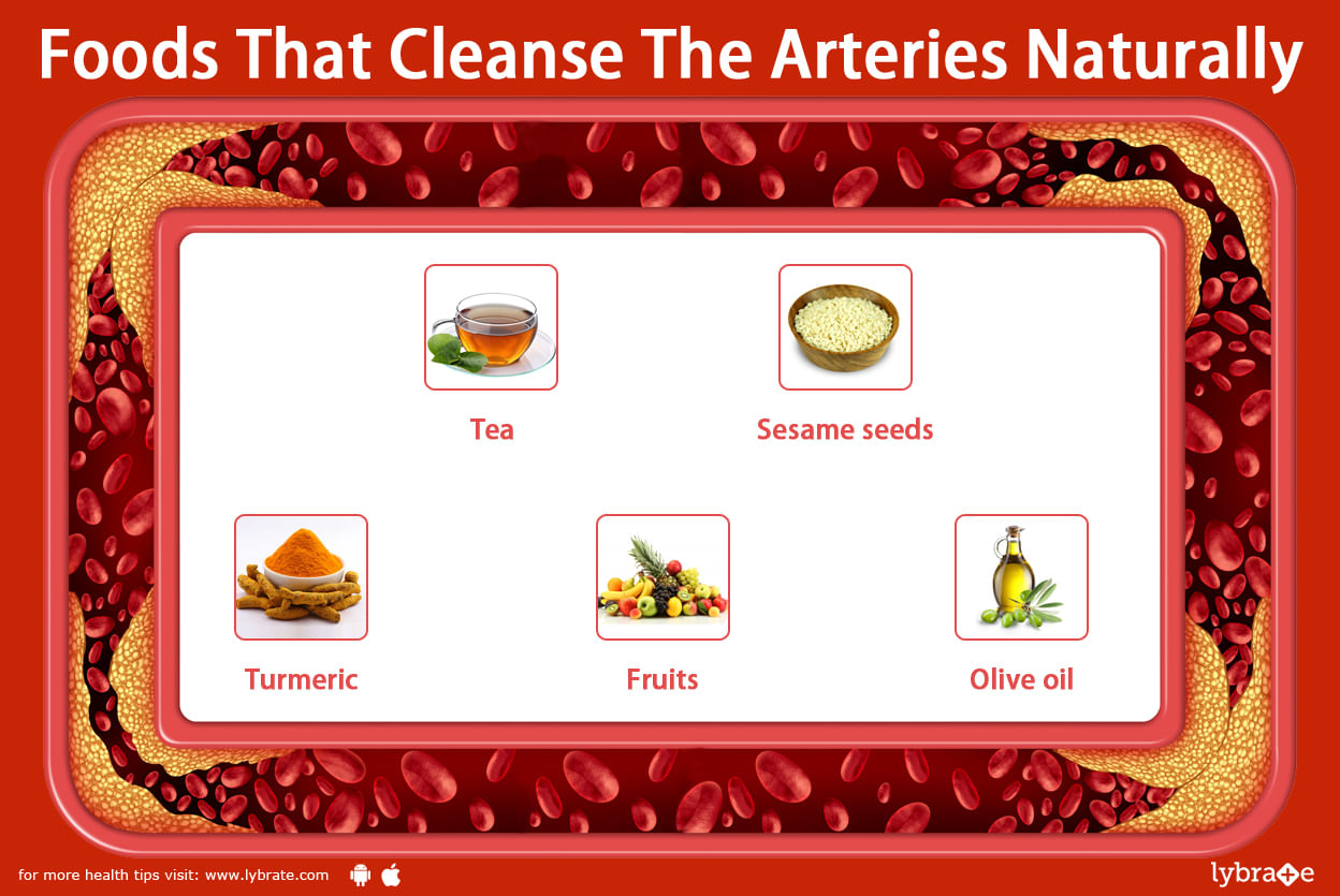
Foods that cleanse the arteries naturally
Arteries are the blood vessels that carry oxygen to various parts of the body. Proper functioning of the arteries is directly related to well functioning organs as without oxygen, no organ can work well. There are many reasons due to which blockages and waste materials are stuck in these blood vessels. It is essential to clean them on regular basis, there are several food items available easily that cleanse the arteries naturally!
1. T...more
Last Updated: 6 years ago• Featured Tip
Share
Bookmark
Report

Blockage in heart is a common term used for narrowing of coronary arteries. Coronary arteries are vessels, which supply blood and thus oxygen and food) to continuously working heart muscles. Heart muscles which are not tired working from the birth till death, however, cannot sustain long without blood supply.
A reduction in blood supply gives rise to ischemia of heart muscles commonly manifested as chest discomfort or angina. A sudden complete shutdown of blood supply leads to heart att...more
A reduction in blood supply gives rise to ischemia of heart muscles commonly manifested as chest discomfort or angina. A sudden complete shutdown of blood supply leads to heart att...more
Last Updated: 8 years ago• Featured Tip
Share
Bookmark
Report

Blockage in heart is a common term used for narrowing of coronary arteries. Coronary arteries are vessels, which supply blood and thus oxygen and food) to continuously working heart muscles. Heart muscles which are not tired working from the birth till death, however, cannot sustain long without blood supply.
A reduction in blood supply gives rise to ischemia of heart muscles commonly manifested as chest discomfort or angina. A sudden complete shutdown of blood supply leads to heart att...more
A reduction in blood supply gives rise to ischemia of heart muscles commonly manifested as chest discomfort or angina. A sudden complete shutdown of blood supply leads to heart att...more

Book appointment with top doctors for femoral artery Image treatment
View fees, clinic timings and reviews

Ask a free question
Get FREE multiple opinions from Doctors
posted anonymously