Get the App
For Doctors
Login/Sign-up

Health Feed
Find Doctors
Health Packages
AllQ&AsTipsQuizzes

Eye Lid Surgery Health Feed
Asked for female, 27 years old from Mumbai
Share
Bookmark
Report


No it will not affect your cararact surgery either, so you can go for it without having any doubt in mind,
9 people found this helpful
Last Updated: 3 years ago• Featured Tip
Share
Bookmark
Report

Masters Of Science In Dietetics And Food...read more
Dietitian/Nutritionist•Mumbai
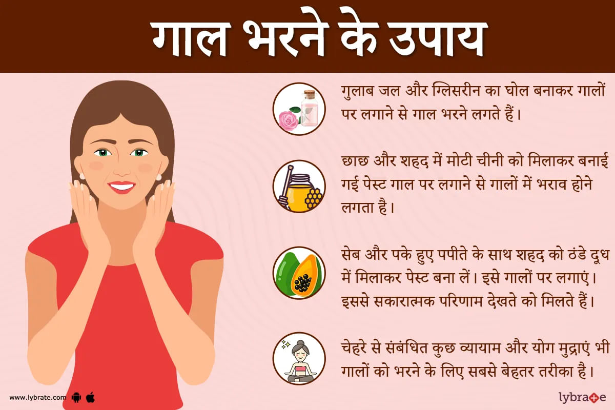
रूप को परिभाषित करने में सुंदर त्वचा टोन के अलावा,चेहरे की विशेषताएं भी महत्वपूर्ण हैं । इसके लिए काम, खान-पान की खराब आदतों, तनाव की वजह से बहुत से लोगों को सूजी हुई आंखों की पलकों, काले घेरों और पिचके और खराब गालों की समस्या हो जाती है। गोल-मटोल गाल चाहे बच्चे हों या बड़े, क्यूटनेस की निशानी है।
भरे हुए गाल से आकर्षक और युवा दिखती हैं। युवा रहने पर में, गालों के नीचे की चमड़े के नीचे की चर्बी एक स्वस्थ, गोल-मटोल और युवा दिखती है। लेकिन उम्र के साथ, गालों के आस-पास के क्षेत्र में त्वच...more
भरे हुए गाल से आकर्षक और युवा दिखती हैं। युवा रहने पर में, गालों के नीचे की चमड़े के नीचे की चर्बी एक स्वस्थ, गोल-मटोल और युवा दिखती है। लेकिन उम्र के साथ, गालों के आस-पास के क्षेत्र में त्वच...more
Last Updated: 3 years ago• Featured Tip
Share
Bookmark
Report

Dr. Ravi thadani
Https://www. Lybrate. Com/delhi/doctor/dr-ravi-thadani-ophthalmologist-1
Mbbs, Ms. In ophthalmology. ;
41 years of experience 500 at clinic 99 online
He is a specialist in treating disorders of eyes with expertise as an ophthalmologist. He is a trained physician, having practised as a botox specialist treating various disorders like periorbital edema, glaucoma and hyperkeratinization of sclera and cornea. With his experience of years an...more
10 people found this helpful
Last Updated: 3 years ago• Featured Tip
Share
Bookmark
Report

1 Dr. Chanchal gadodiya
Https://www. Lybrate. Com/pune/doctor/dr-chanchal-gadodiya-ophthalmologist
M. B. B. S, ms, d. N. B, fellowship in paediatric opth & strabismology
An eye doctor specializing in low vision eyes lasik, and laser eye surgeon, vitreoretinal, cataract, and glaucoma surgeon oculoplasty and cornea transplant surgeon a pediatric strabismus specialist with a master of science from b y l nair hospital in mumbai and a bachelor of medicine and...more
Last Updated: 3 years ago• Featured Tip
Share
Bookmark
Report

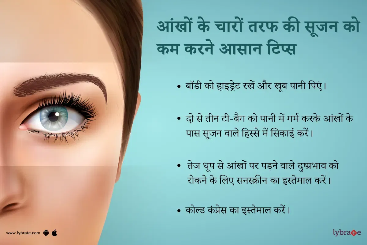
हम अपने शरीर को सुडौल और चेहरे को सुंदर बनाए रखने के लिए तरह तरह के उपाय करते रहते हैं। त्वचा की देखभाल के ढेरों नुस्खें अपनाते हैं।पर कई बार आपके चांद से चेहरे पर सूजी हुई आंखें दाग लगा देती हैं। आंखों के नीचे की सूजन चेहरे की ताज़गी पर सवाल खड़े कर देती है। ये दरअसल रोजाना की थकान और नींद की कमी से हो सकता है।इससे छुटकारा पाने के लिए आप पर्याप्त मात्रा में आराम करने से काफी फर्क पड़ता है।पर कई बार सिर्फ इतना ही काफी नहीं होता। आपकी आंखों के आसपास की त्वचा अत्यधिक संवेदनशील होती है जिसपर छोटे प...more
1243 people found this helpful
Health Query
Share
Bookmark
Report

Generally lasik surgery doesn't cause sight back if your having blindness from birth then it's not possible, but if retina is given then there is every chance of getting back your eye sight.
154 people found this helpful
Asked for male, 25 years old from Saharanpur
Share
Bookmark
Report

Hello. First of all, -4.5 dioptre of refractory error (glasses) do not result from excessive computer work. It is just simple myopia. Yes you can go for lasik or other more advanced similar laser procedures. No need to worry. After surgery eye sight will not be affected due to your longer screen time of 10-12 hours. But you will have some dry eye problem after lasik, so you will have to take more care due to your job profile. Certain drops post surgery along with certain eye exercises, intermitt...more
Health Query
Share
Bookmark
Report

Yes u can go for lasik,, there is no such risk,, it costs around 40k to 50k per eyes may be higher in some places
Asked for Male, 22 years old from Pune
Share
Bookmark
Report


Regarding how long you will have to take the medications- you will need to speak with the Dr. who prescribed them since he would have examined and prescribed accordingly.
And just for 2 days there won't be any effects on your lasik surgery outcome do continue your lasik medications as advised. Do make sure you have informed your own lasik surgeon also about these medications.
And just for 2 days there won't be any effects on your lasik surgery outcome do continue your lasik medications as advised. Do make sure you have informed your own lasik surgeon also about these medications.
Asked for male, 22 years old from Kannur
Share
Bookmark
Report

It is because of the progress of the disease
It can be stopped by homoeopathic treatment.
You can consult me at lybrate.
It can be stopped by homoeopathic treatment.
You can consult me at lybrate.
3 people found this helpful

Book appointment with top doctors for Eye Lid Surgery treatment
View fees, clinic timings and reviews

Ask a free question
Get FREE multiple opinions from Doctors
posted anonymously