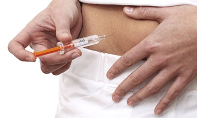
Chorionic Villus Sampling Test
Am 30 years female. I have been married for 5 years. In these years I had 2 miscarriages one in 2022 and another in 2023 ...


Ask Free Question
Is yours is a consanguinity, better to have chorionic villus sampling or chromosomal analysis to ruleout problem.


Down syndrome adult 23 years old After a period of 2-3 months, he gets cough in chest and nausea and vomiting simultaneo ...

Ask Free Question
Amniocentesis is usually performed between week 16 and week 18, though it can be performed as soon as week 15 or as late as week 20. Amnio looks for several hundred genetic diseases, including Down syndrome, Tay-Sachs and sickle cell anemia. Unlike chorionic villus sampling (CVS), amnio can also rule out neural tube defects such as spina bifida. It does not, however, detect every kind of abnormality, including cleft lip or palate. And it can't determine the severity of the problem. you may need a detailed discussion for further consultation.

I and my partner want to get married but we both have thalassemia minor. We are facing a lot of objections from our fami ...

Ask Free Question
Yes there is 25% possibility that the kid will be thalassemia major. But you can do following test during pregnancy to find out if your baby is at risk for thalassemia, you can have prenatal tests during pregancy to see if he has the condition. Prenatal tests are tests you get during pregnancy to see how you and your baby are doing. 1. Chorionic villus sampling (also called cvs). This test checks tissue from the placenta to see if your baby has a genetic condition. You can get cvs at 10 to 13 weeks of pregnancy. 2. Amniocentesis (also called amnio). This test takes some amniotic fluid from around your baby in the uterus. The test checks for birth defects and genetic conditions in your baby. You can get this test at 15 to 20 weeks of pregnancy.
Hi Sir, me and my girlfriend both are having thalassemia carrier. We want to marry now. Is there any problem in our sexu ...

Ask Free Question
No. Don't worry. Accuracy of the CVS Test. Chorionic villus sampling is more than 99 percent accurate when it comes to diagnosing chromosomal results, such as Down syndrome. However, there's a sliver of a chance for a false positive—when the test comes back indicating a genetic problem, but in reality, the baby is developing normally.
What cause abnormal babies development in mother womb and in which month it can be known during pregnancy. ...

Ask Free Question
An estimated 303 000 newborns die within 4 weeks of birth every year, worldwide, due to congenital anomalies. Congenital anomalies can contribute to long-term disability, which may have significant impacts on individuals, families, health-care systems, and societies. The most common, severe congenital anomalies are heart defects, neural tube defects and Down syndrome. Although congenital anomalies may be the result of one or more genetic, infectious, nutritional or environmental factors, it is often difficult to identify the exact causes. Some congenital anomalies can be prevented. Vaccination, adequate intake of folic acid or iodine through fortification of staple foods or supplementation, and adequate antenatal care are just 3 examples of prevention methods. Causes and risk factors Although approximately 50% of all congenital anomalies cannot be linked to a specific cause, there are some known genetic, environmental and other causes or risk factors. Genetic factors Genes play an important role in many congenital anomalies. This might be through inherited genes that code for an anomaly, or resulting from sudden changes in genes known as mutations. Consanguinity (when parents are related by blood) also increases the prevalence of rare genetic congenital anomalies and nearly doubles the risk for neonatal and childhood death, intellectual disability and other anomalies. Some ethnic communities (such as Ashkenazi Jews or Finns) have a comparatively high prevalence of rare genetic mutations such as Cystic Fibrosis and Haemophilia C. Socioeconomic and demographic factors Low-income may be an indirect determinant of congenital anomalies, with a higher frequency among resource-constrained families and countries. It is estimated that about 94% of severe congenital anomalies occur in low- and middle-income countries. An indirect determinant, this higher risk relates to a possible lack of access to sufficient, nutritious foods by pregnant women, an increased exposure to agents or factors such as infection and alcohol, or poorer access to healthcare and screening. Factors often associated with lower-income may induce or increase the incidence of abnormal prenatal development. Maternal age is also a risk factor for abnormal intrauterine fetal development. Advanced maternal age increases the risk of chromosomal abnormalities, including Down syndrome. Environmental factors Maternal exposure to certain pesticides and other chemicals, as well as certain medications, alcohol, tobacco and radiation during pregnancy, may increase the risk of having a fetus or neonate affected by congenital anomalies. Working or living near, or in, waste sites, smelters or mines may also be a risk factor, particularly if the mother is exposed to other environmental risk factors or nutritional deficiencies. Infections Maternal infections such as syphilis and rubella are a significant cause of congenital anomalies in low- and middle-income countries. More recently, the effect of in utero exposure to Zika virus on the developing fetus has been reported. In 2015, Brazil detected cases of Zika virus and a spatio-temporally associated increase in microcephaly. By 2016, Brazil reported that of 4180 suspected cases of microcephaly, 270 were confirmed, 462 were discarded and 3448 are still under investigation. This is compared to an average of 163 microcephaly cases recorded nationwide per year. With 6 of the 270 confirmed cases of microcephaly showing evidence of Zika infection, health authorities and agencies are investigating and conducting comprehensive research to confirm a causal link. Following the Zika outbreak in French Polynesia, health authorities reported an unusual increase in the number of congenital malformations in babies born between March 2014 and May 2015. Maternal nutritional status Maternal folate insufficiency increases the risk of having a baby with a neural tube defect while excessive vitamin A intake may affect the normal development of an embryo or fetus. Detection Health care before and around the time of conception (preconception and peri-conception) includes basic reproductive health practices, as well as medical genetic screening and counselling. Screening can be conducted during the 3 periods listed: Preconception screening can be useful to identify those at risk for specific disorders or at risk of passing a disorder onto their children. Screening includes obtaining family histories and carrier screening, and is particularly valuable in countries where consanguineous marriage is common. Peri-conception screening: maternal characteristics may increase risk, and screening results should be used to offer appropriate care, according to risk. This may include screening for young or advanced maternal age, as well as screening for use of alcohol, tobacco or other risks. Ultrasound can be used to screen for Down syndrome and major structural abnormalities during the first trimester, and for severe fetal anomalies during the second trimester. Maternal blood can be screened for placental markers to aid in prediction of risk of chromosomal abnormalities or neural tube defects, or for free fetal DNA to screen for many chromosomal abnormalities. Diagnostic tests such as chorionic villus sampling and amniocentesis can be used to diagnose chromosomal abnormalities and infections in women at high risk. Neonatal screening includes clinical examination and screening for disorders of the blood, metabolism and hormone production. Screening for deafness and heart defects, as well as early detection of congenital anomalies, can facilitate life-saving treatments and prevent progression towards some physical, intellectual, visual, or auditory disabilities. In some countries, babies are routinely screened for abnormalities of the thyroid or adrenal glands before discharge from the maternity unit.

My girlfriend and me both have sickle-cell carrier in our blood. Is there any (other than IVF technique, adopting )way t ...

Ask Free Question
When both partners are sickle cell carrier then each baby has 1 in 2 risk of being a carrier ,1 in4 risk of having sickle cell anaemia and 1 in4 chance of not being a carrier Ivf is not required ,can go for normal conception ,but prenatal testing - either amniocentesis or chorionic villus sampling should be done at around 10 weeks of pregnancy to find out the sickle cell status of baby.

Me and my spouse are thalassemia minor Our one daughter is also thalassemia minor, We lost our one daughter, she was tha ...
Ask Free Question
As you both are thalassemia minor pt and have lost a child to thalassemia major, your wife should undergo chorionic villus sampling test at 10-12 weeks of pregnancy. This is an invasive test in which tissue from placenta is taken and fetal DNA analysis done. If the fetus is having thalassemia minor you can continue the pregnancy, if major you should terminate the pregnancy.

I am a thalassemia minor and my partner is also minor. What should we hav to do for healthy baby? ...

Ask Free Question
If both you and your partner are thalassaemia minor then there is a 25% chance (1 in 4 chance) that your baby could be affected by thalassemia major which is a debilitating disease. You should meet a genetic medicine specialist for detailed counselling. If you are already pregnant, then there are some ways that doctors can find out whether the unborn child has thalassemia major. The techniques used are: chorionic villus sampling or by fetal blood sampling. Both tests are conducted under ultrasound guidance. Chorionic villus sampling involves obtaining some cells from the placenta for testing. This is done during the 10th to 12th week of pregnancy. Fetal blood sampling involves obtaining a small amount of fetal blood from the umbilical cord for testing. This procedure is done at about 18 - 20 weeks of pregnancy.
Last year, in April I had a miscarriage in 3 months of pregnancy. Then doctor test the product and it was Turner syndrom ...

Ask Free Question
Dear lybrate user, to avoid turner syndrome which obstruct development in female, and may cause, premature ovarian failure, relax! * take, homoeo-medicine* @ sepia 30-6 pills, thrice a day. @ ova tosta 3x -4 tabs, thrice a day. * go for meditation, to reduce your stress and anxiety and generate strong will power that you will wine d game. * do not exert and take, maximum rest in the present condition. * take, easily digestible diet, follow the instruction of your gynae and act accordingly. All d best.