Get the App
For Doctors
Login/Sign-up

Health Feed
Find Doctors
Health Packages

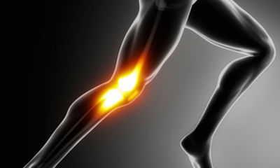
Patellar Health Feed
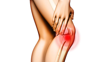
Asked for male, 25 years old from Kangra
Share
Bookmark
Report

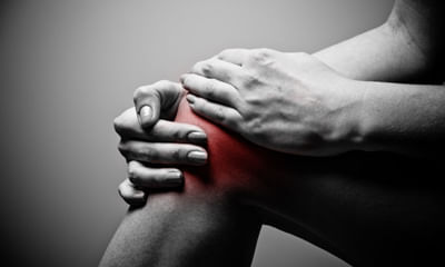
Hello according to your report your posterior meniscus is tear. You have to need knee braces to maintain knee in extension.
Knee braces sote time v laga k hi sona.
While walking donot give full weight.
In physiotherapy session you take ultrasonic therapy for 12 mints on posteriorly of knee.
Ift therapy for 15 mints posteriorly knee.
In home you take ice or cold pack for 20 mints on back of knee.
Take rest for 4 weeks.
Because ligaments tear take 6-8 of healing time so...more
Health Query
Share
Bookmark
Report

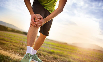
Sports Taping- stretch the tape from both ends and apply on the affected area
Contrast Fomentation (Hot and Cold).
Contrast Fomentation (Hot and Cold).
45 people found this helpful
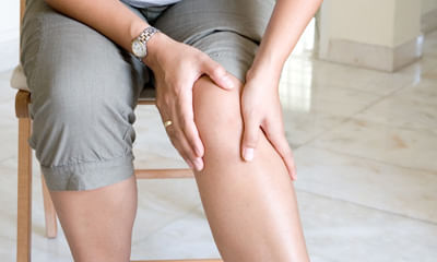
Asked for male, 66 years old from Calicut
Share
Bookmark
Report
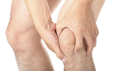
Chiropractic adjustment will help.
Avoid Squatting-
Avoid sitting Cross legged.
Avoid Squatting-
Avoid sitting Cross legged.
102 people found this helpful
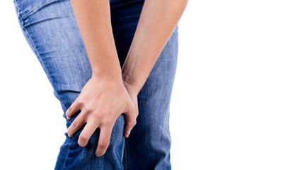
Asked for male, 33 years old from Chennai
Share
Bookmark
Report
Hi lybrate-user U can go in for Physiotherapy treatment For the pain and once the pain is gone you learn the Quadriceps and Hamstring strengthening exs and do them regulatly.
24 people found this helpful
Health Query
Share
Bookmark
Report
Over weight case, don't advise, take sky fruit, cow urine cap, org wheat grass powder, curcumin droops, drink 30 ml extra virgin coconut oil, seabuckthoren juice, put almond oil into both nostel at bedtime, take chamomile black tea, brahami gold tab daily, may contact for any assistance.
22 people found this helpful
Asked for male, 32 years old from Jaipur
Share
Bookmark
Report
This is a general strain due to excessive walking or standing and for this you can follow these measures: one keep a pillow right under the knee while sleeping, next is you can keep ice in the painful area for about 5--10 minutes. One time you can do hot water fermentation that would help to reduce the muscle strain
arthritic Pain:Ice therapy would definitely help to reduce the inflammation. We also advise you to use knee cap which would help to prevent the knee from damaging further and als...more
arthritic Pain:Ice therapy would definitely help to reduce the inflammation. We also advise you to use knee cap which would help to prevent the knee from damaging further and als...more
86 people found this helpful
Health Query
Share
Bookmark
Report

Dear ,
greetings
description tells me that you might have sub-clinical infection. As you are off antibiotics now, we need to see how your feels after 2-3 wks. Repeat your crp and esr levels. 20 days post surgery there should not be any other cause of fever even though its mild. Swelling will take long time to around 6 months or so. Your doctor may advice culture from knee fluid once your are off antibiotics for about 2 wks. Was the second mri with contrast?
You have more pain in bone...more
Health Query
Share
Bookmark
Report
The typical symptoms of a posterior cruciate ligament injury are:
•pain with swelling that occurs steadily and quickly after the injury.
•swelling that makes the knee stiff and may cause a limp.
•difficulty walking.
•the knee feels unstable, like it may "give out"
the posterior cruciate ligament (pcl) is the strongest ligament in the knee. It extends from the top-rear surface of the tibia (bone between the knee and ankle) to the bottom-front surface of the femur (bone that ex...more
50 people found this helpful
Asked for male, 23 years old from Delhi
Share
Bookmark
Report
Send your mri photo.
What problm you r facing.
Can take tab tendomac 1bd for 6 months.
Counslt to physiotherpist.
Do exercises advised by physio.
Apply ice pack n use knee brace.
What problm you r facing.
Can take tab tendomac 1bd for 6 months.
Counslt to physiotherpist.
Do exercises advised by physio.
Apply ice pack n use knee brace.
44 people found this helpful
Asked for male, 23 years old from Hyderabad
Share
Bookmark
Report

The knee-jerk reflex, also known as the patellar reflex, is a simple reflex that causes the contraction of the quadriceps muscle when the patellar tendon is stretched. Let's have a detailed discussion for better advice and medication plan.
30 people found this helpful

Book appointment with top doctors for Patellar treatment
View fees, clinic timings and reviews

Ask a free question
Get FREE multiple opinions from Doctors
posted anonymously