Lybrate Logo
Get the App
For Doctors
Login/Sign-up
Book Appointment
Treatment
Ask a Question
Plan my Surgery
Health Feed
tab_logos
About
tab_logos
Health Feed
tab_logos
Find Doctors
tab_logos
Health Packages
Facebook Icon
Twitter Icon
Telegram Icon
Linkedin Icon

Kniest Dysplasia Questions

Health Query
Share
Bookmark
Report
Hip dysplasia has 3 grade , what about his norbeys angle ?how is his muscle mass of leg? How is nerve sensation. If u put some x rays and dog photo it will help me to decide line of treatment.
Asked for male, 39 years old from Mumbai
Share
Bookmark
Report

Dear Sir/Madam, I had suffered from hepatitis ‘E’ along with leptospirosis infection in the year 2011. Post recovery I was feeling very weak along-with very much weakness in the body. As the passing of the time I usually felt good and my weight has also been increased. Doctor told to make to take everything in the diet. But from the last 3-4 years I am experiencing lots of changes in my body like poor digestion, excessive gas, bloating, indigestion, loose stool with mucus, 5-6 times visiting the toilet, fatigue,lethargy, weakness in the body, irritability,fear, nervousness,easily stressed out. Not able to get sound sleep at nights. I got a very weak immune system also. Visited lots of doctors here in Mumbai. Some doctors says I got physiological problems. Some says I have gastritis, some says I have IBS, some says that I must be having amoebiasis or H.Pylori infections. Some says that there is inflammation in the intestines and my intestines are weak. I am not getting proper solution to my problem. At times I am feeling very much depressed. I do not know what to do. I am feeling hopeless. I do not know what had happened to my body. Nobody able to properly diagnose the problem which I am having. I do not have any bad habits. I do not smoke, drink etc. But still I am so much problem in my body. Further, below are the test performed in order to rule out. OGD SCOPY + is can + biopsy PERFORMED OGD Scopy was considered to evaluate exact etiology of patients symptoms of Upper G.I.Tract. OGD Scopy revealed mild lax cardia with frank reflux. Stomach showed erosive gastritis with atrophic antral mucosa. Erosions were seen in the fundus and antrum. Chronic duodenitis was seen in the duodenal bulb and proximal duodenum. Biopsy was taken from antrum for H.Pylori. Comments: Endoscopic findings are suggestive of mild GERD with erosive gastritis with atrophic antral mucosa and chronic duodenitis. Patient will require dietary and lifestyle modifications apart from supportive medication for the same. Patient will require clinical follow up after 1 month. Biopsy confirmed for H.Pylori. ILEO-COLONOSCOPY+ is can+POLYPECTOMY PERFORMED Ileo-Colonoscopy was considered to evaluate exact etiology for patients complaints of 3-4 semi solids with blood streak stools. Adequate bowel preparation was done. Scope was passed up to terminal ileum, which essentially appeared normal. Multiple biopsies were taken from colon and sent for HPE. Left sided colon appeared spastic. Erosive proctitis was seen in rectum. 2 polyps were noted on the stapled line on previously done hemorrhoidectomy which was sent for histopathology. Comments: Ileo-colonoscopy findings are suggestive of irritable spastic left bowel with erosive proctitis and 2 polyps at the stapled line which can explain patients symptoms of blood streak stools. Biopsy material may provide additional information for further management. Patient will require reassurance apart from supportive medical care. Patient will require clinical follow up after 1 month. HISTOLOGY REPORT Specimen:1.Biopsies from the colon 2. Polyp from the rectum Gross:1. Three tiny bits aggregate to 0.4 cm. 2. Few polypoid grayish bits aggregate to 0.4 cm. Microscopic: 1. The colonic mucosa reveals orderly tubules. The lamina propria contains a mononuclear inflammatory infiltrate admixed with eosinophil and a focal lymphoid aggregate. 2. The polypoid bits are focally covered by a colonic mucosa. The lamina propria shows congested blood vessels and a moderate mononuclear inflammatory infiltrate admixed with neutrophil few eosinophil. No adenomatous change is seen. There is no evidence of dysplasia or malignancy. Diagnosis.1. COLON – MILD colitis 2. RECTUM – INFLAMMATORY POLYP.

Profile Image

Bachelor of Unani Medicine and Surgery (...read more

Ayurveda•Kanpur
banner-image
sootshekhar ras 125 mg twice a day
panchsakar churna 50 gm + avipattikar churna 40 gm + karpad bhasm 10 gm + shankh bhasm 5 gm SAB KO MILAKAR 5 GM twice a day
133 people found this helpful
Asked for male, 39 years old from Mumbai
Share
Bookmark
Report

Dear Sir/Madam, I had suffered from hepatitis ‘E’ along with leptospirosis infection in the year 2011. Post recovery I was feeling very weak along-with very much weakness in the body. As the passing of the time I usually felt good and my weight has also been increased. Doctor told to make to take everything in the diet. But from the last 3-4 years I am experiencing lots of changes in my body like poor digestion, excessive gas, bloating, indigestion, loose stool with mucus, 5-6 times visiting the toilet, fatigue,lethargy, weakness in the body, irritability,fear, nervousness,easily stressed out. Not able to get sound sleep at nights. I got a very weak immune system also. Visited lots of doctors here in Mumbai. Some doctors says I got physiological problems. Some says I have gastritis, some says I have IBS, some says that I must be having amoebiasis or H.Pylori infections. Some says that there is inflammation in the intestines and my intestines are weak. I am not getting proper solution to my problem. At times I am feeling very much depressed. I do not know what to do. I am feeling hopeless. I do not know what had happened to my body. Nobody able to properly diagnose the problem which I am having. I do not have any bad habits. I do not smoke, drink etc. But still I am so much problem in my body. Further, below are the test performed in order to rule out. OGD SCOPY + is can + biopsy PERFORMED OGD Scopy was considered to evaluate exact etiology of patients symptoms of Upper G.I.Tract. OGD Scopy revealed mild lax cardia with frank reflux. Stomach showed erosive gastritis with atrophic antral mucosa. Erosions were seen in the fundus and antrum. Chronic duodenitis was seen in the duodenal bulb and proximal duodenum. Biopsy was taken from antrum for H.Pylori. Comments: Endoscopic findings are suggestive of mild GERD with erosive gastritis with atrophic antral mucosa and chronic duodenitis. Patient will require dietary and lifestyle modifications apart from supportive medication for the same. Patient will require clinical follow up after 1 month. Biopsy confirmed for H.Pylori. ILEO-COLONOSCOPY+ is can+POLYPECTOMY PERFORMED Ileo-Colonoscopy was considered to evaluate exact etiology for patients complaints of 3-4 semi solids with blood streak stools. Adequate bowel preparation was done. Scope was passed up to terminal ileum, which essentially appeared normal. Multiple biopsies were taken from colon and sent for HPE. Left sided colon appeared spastic. Erosive proctitis was seen in rectum. 2 polyps were noted on the stapled line on previously done hemorrhoidectomy which was sent for histopathology. Comments: Ileo-colonoscopy findings are suggestive of irritable spastic left bowel with erosive proctitis and 2 polyps at the stapled line which can explain patients symptoms of blood streak stools. Biopsy material may provide additional information for further management. Patient will require reassurance apart from supportive medical care. Patient will require clinical follow up after 1 month. HISTOLOGY REPORT Specimen:1.Biopsies from the colon 2. Polyp from the rectum Gross:1. Three tiny bits aggregate to 0.4 cm. 2. Few polypoid grayish bits aggregate to 0.4 cm. Microscopic: 1. The colonic mucosa reveals orderly tubules. The lamina propria contains a mononuclear inflammatory infiltrate admixed with eosinophil and a focal lymphoid aggregate. 2. The polypoid bits are focally covered by a colonic mucosa. The lamina propria shows congested blood vessels and a moderate mononuclear inflammatory infiltrate admixed with neutrophil few eosinophil. No adenomatous change is seen. There is no evidence of dysplasia or malignancy. Diagnosis.1. COLON – MILD colitis 2. RECTUM – INFLAMMATORY POLYP.

Profile Image

Bachelor of Unani Medicine and Surgery (...read more

Ayurveda•Kanpur
banner-image
sootshekhar ras 125 mg twice a day
panchsakar churna 50 gm + avipattikar churna 40 gm + karpad bhasm 10 gm + shankh bhasm 5 gm SAB KO MILAKAR 5 GM twice a day
194 people found this helpful
Asked for male, 26 years old from Siliguri
Share
Bookmark
Report
Profile Image

MD-Ophthalmology

Ophthalmologist•Srinagar
It is called as naevus.
If it is increasing in size or changes color, may signify dysplasia (early malignant changes. Kindly consult ophthalmologist, and get it removed.
They will then do a histopathological test on tissue for diagnosis.
Yes, it can be removed and need urgent attention.
Asked for female, 34 years old from Srinagar
Share
Bookmark
Report
Profile Image

M.Ch - Surgical Gastroenterology/G.I. Su...read more

Gastroenterologist•Calicut
banner-image
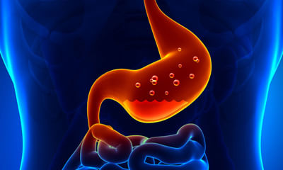
Gallstones if asymptomatic need no treatment, that means no Medicine or surgery. If symptoms are there that is pain in Right upper abdomen, dysplasia, retrostenal pain, pain radiating to back, fever with chills or jaundice you require treatment and that is surgery. Key hole surgery laproscopic cholecystectomy.
48 people found this helpful
Asked for Male, 28 years old from Jhajjar
Share
Bookmark
Report
Profile Image

MBBS, Basic Life Support (B.L.S), Advanc...read more

General Physician•Delhi
Dysplasia is an abnormal growth or development of cells and ulcerative colitis may be due to inflammatory disease conditions. With early identification, treatment and consistent follow up it can be cured. Let's have a detailed discussion for better advice and healthy lifestyle.
67 people found this helpful
Asked for male, 41 years old from Vadodara
Share
Bookmark
Report
Profile Image

MDS - Oral & Maxillofacial Surgery

Dentist•Chennai
banner-image
1)Replacement of amalgam restorations
2)Withdrawal of the drug
3)Oral lichen lesions in chronic in graft versus host disease are usually managed with local corticosteroids or other drugs such as tacrolimus.
4)As in OLP the question arises whether one or all types of oral lichenoid lesions are to be considered a potentially malignant disorder
In the absence of known etiological factors, the taking of a biopsy should be considered, particularly in case of a non-reticular lesion, in...more
662 people found this helpful
Health Query
Share
Bookmark
Report
Profile Image

C.S.C, D.C.H, M.B.B.S

General Physician•Alappuzha
banner-image
In some people both kidneys can be affected by congenital dysplasia, and there may be kidney failure. ... An infection in a kidney can cause it to shrink. Normally kidney infections do not cause permanent damage to a kidney, or leave a small scarred area in the kidney.Depending on the underlying cause, some types of kidney disease can be treated. Often, though, chronic kidney disease has no cure. ... If your kidneys become severely damaged, you may need treatment for end-stage kidney disease.
Asked for female, 28 years old from Tumkur
Share
Bookmark
Report
Profile Image

MDS - Oral & Maxillofacial Surgery

Dentist•Chennai
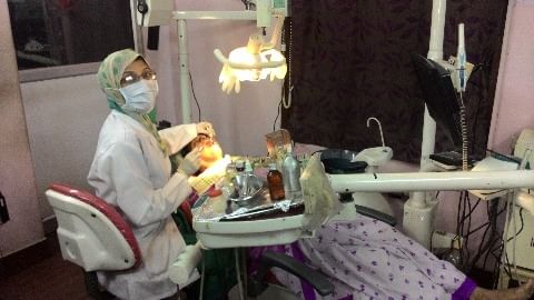
ORAL LICHENOID LESIONS
Four types of oral lichenoid lesions (OLLs) can be distinguished, being
1) Amalgam restoration, topographically associated lesions,
2) Drug related lichenoid lesions,
3) Lichenoid lesions in chronic graft versus host disease (cGVHD), and
4) Unclassified (e.g. erythematous changes limited to the gingiva without signs of “classic” oral lichen planus elsewhere in the oral cavity, or lesions that have a lichen planus like aspect but that lack one or mo...more
book_appt_icon
Book appointment with top doctors for Kniest Dysplasia treatment
View fees, clinic timings and reviews
chat_icon

Ask a free question

Get FREE multiple opinions from Doctors

posted anonymously
Pristyn Care Banner