Get the App
For Doctors
Login/Sign-up

About
Health Feed
Find Doctors
Health Packages

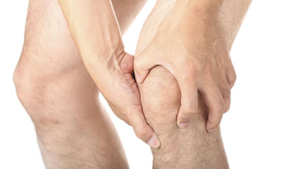
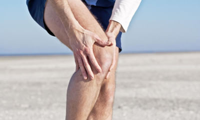
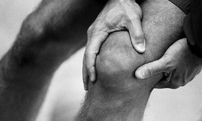
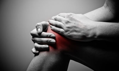
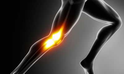
Knee Ligaments Image Health Feed
Asked for male, 21 years old from Abohar
Share
Bookmark
Report
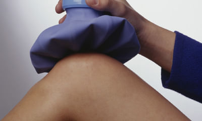
Avoid weight bearing if knee is painful. Walk with the help of elbow crutches in both hands. Apply elastocrepe bandage and ice over knee. Start physiotherapy. Consult specialist in arthroscopy and sports medicine for further treatment.
235 people found this helpful
Asked for male, 19 years old from Abohar
Share
Bookmark
Report
Hi lybrate-user. I'm sure the ligament injury has been picked up during the mri scan. Please see a good arthroscopists. Meantime take some anti inflammatory medicines to curb your pain and swelling. Revert back for help.
174 people found this helpful
Asked for male, 23 years old from Delhi
Share
Bookmark
Report
Send your mri photo.
What problm you r facing.
Can take tab tendomac 1bd for 6 months.
Counslt to physiotherpist.
Do exercises advised by physio.
Apply ice pack n use knee brace.
What problm you r facing.
Can take tab tendomac 1bd for 6 months.
Counslt to physiotherpist.
Do exercises advised by physio.
Apply ice pack n use knee brace.
44 people found this helpful
Asked for female, 20 years old from Jabalpur
Share
Bookmark
Report

Take rest. Homoeopathy has good treatment for your problem, without causing adverse effects. To start with, take homoeopathic medicine arnica 200 daily in the morning and in the evening and give feedback after 4 days.
Health Query
Share
Bookmark
Report
Its a small procedure where the surgeon will put a small incision to insert a camera to diagnose the condition. Cost may vary from hospitals.
Asked for male, 25 years old from Chandigarh
Share
Bookmark
Report
Innitially apply ice and crepe bandage to affected knee. You will require static quadriceps and hamstring exercises. If no relief in 3 weeks and pain continues or you r not comfortable with your knee, you will require to get mri of the affected knee.
153 people found this helpful
Health Query
Share
Bookmark
Report
Hi
for initial treatment of a pcl injury,
1) protecting the knee from further injury
2) resting the knee
3) icing the knee for short periods with cold packs
4) compressing the knee gently, such as with an elastic bandage
5) elevating the knee
for detail treatment plan visit physiotherapist or orthopaedic doctor.
Best wishes.
Asked for male, 27 years old from Kota
Share
Bookmark
Report
It must be investigated
Get a MRI of the knee.
You would probably need arthroscopic knee surgery.
It is a very safe procedure with uniformly good results.
Don?t ignore it lest it become beginning of a bigger problem
Why not discuss with me in a video conference? ( Facility provided by lybrate.com)
Get a MRI of the knee.
You would probably need arthroscopic knee surgery.
It is a very safe procedure with uniformly good results.
Don?t ignore it lest it become beginning of a bigger problem
Why not discuss with me in a video conference? ( Facility provided by lybrate.com)
421 people found this helpful
Health Query
Share
Bookmark
Report

Calcium and protein required Wax bath therapy for 20 days Constitution with orthopaedic arthroscopic surgeon.
13 people found this helpful
Health Query
Share
Bookmark
Report
Complete recovery may not possible,,u can dance but not now,,if u feel pain after dancing then u to stop dancing,

Book appointment with top doctors for Knee Ligaments Image treatment
View fees, clinic timings and reviews

Ask a free question
Get FREE multiple opinions from Doctors
posted anonymously