Get the App
For Doctors
Login/Sign-up

About
Health Feed
Find Doctors
Health Packages

Foot Ligaments Image Health Feed
Asked for female, 67 years old from Panchkula
Share
Bookmark
Report

Please send me ultrasound reports meanwhile take plenty of water daily take sy neeri by aimil pharma 20 ml in 200 ml water three times a day for 3 weeks.
27 people found this helpful
Asked for female, 42 years old from Hisar
Share
Bookmark
Report

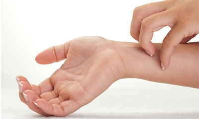
It can be because of dermatitis/ eczema or allergy or dryness or fungal infection or psoaiasis etc. I need details of case n preferably pics of affected area. In the meanwhile follow this
moisturize frequently.
Avoid sudden changes in temperature or humidity.
Avoid sweating or overheating.
Reduce stress.
Avoid scratchy materials, such as wool directly in contact with skin.
Avoid harsh soaps, detergents, and solvents.
Be aware of any foods that may cause an outbreak an...more
moisturize frequently.
Avoid sudden changes in temperature or humidity.
Avoid sweating or overheating.
Reduce stress.
Avoid scratchy materials, such as wool directly in contact with skin.
Avoid harsh soaps, detergents, and solvents.
Be aware of any foods that may cause an outbreak an...more
14 people found this helpful
Asked for male, 21 years old from Kishanganj
Share
Bookmark
Report

Hello lybrate user,
you will need a complete anterior cruciate ligament repair / reconstruction by arthroscopic techniques after you have regained normal knee joint movements.
This is a special surgery so consult a specialist for details. Follow up.
God bless you.
you will need a complete anterior cruciate ligament repair / reconstruction by arthroscopic techniques after you have regained normal knee joint movements.
This is a special surgery so consult a specialist for details. Follow up.
God bless you.
324 people found this helpful
Health Query
Share
Bookmark
Report

Hi thanks for your query and welcome to lybrate. I am dr akshay from fortis hospital, new delhi. Can you please tell me which ligament are you talking about? it will be helpful if you can upload mri ( if done) images and report along with doctor' s prescription papers, so that I can see and advise you further on it. Do not hesitate to contact me if you need any further assistance.
Thanks & regards
Dr Akshay Kumar Saxena
Thanks & regards
Dr Akshay Kumar Saxena
295 people found this helpful
Health Query
Share
Bookmark
Report

Dear Lybrate user
During injury you may feel a pop in your knee or hear a distinct popping sound, followed by which you end up crashing on the floor if you fail to find any support to hold on to.A quick onset of unbearable leg pain follows the pop. It can be a burning sensation or a shooting pain that makes your nerves from the knee to the hip go crazy. At this stage you would be unable to walk, stand or apply any kind of pressure on your kneeYou will see that your knee has swollen t...more
During injury you may feel a pop in your knee or hear a distinct popping sound, followed by which you end up crashing on the floor if you fail to find any support to hold on to.A quick onset of unbearable leg pain follows the pop. It can be a burning sensation or a shooting pain that makes your nerves from the knee to the hip go crazy. At this stage you would be unable to walk, stand or apply any kind of pressure on your kneeYou will see that your knee has swollen t...more
86 people found this helpful
Asked for male, 23 years old from Delhi
Share
Bookmark
Report

if the ligament injury had been a complete tear then it wouldnt heal spontaneously and that would give rise to recurrent instability symptoms. if you have pain and a sense of giving way in the ankle then you would need a proper orthopaedic examination. you may be advised a course of physio or surgery depending on the examination findings and imaging results
183 people found this helpful
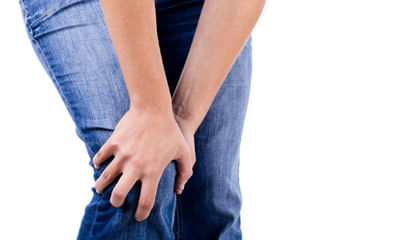
Asked for female, 54 years old from Kolkata
Share
Bookmark
Report
Hi thanks for your query and welcome to lybrate. I am Dr. Akshay from fortis hospital, new delhi. You are around 51 years of age, and pain is towards inner knee aspect.
Anyway is it possible for you to send me a clinical picture of your knee and an x ray if you have already done it? also tell me whether there is any associated swelling, skin redness or local warmth over the skin?
what medicines do you generally take for other conditions? my advice to you till the time I can see your inve...more
Asked for male, 23 years old from Delhi
Share
Bookmark
Report
Send your mri photo.
What problm you r facing.
Can take tab tendomac 1bd for 6 months.
Counslt to physiotherpist.
Do exercises advised by physio.
Apply ice pack n use knee brace.
What problm you r facing.
Can take tab tendomac 1bd for 6 months.
Counslt to physiotherpist.
Do exercises advised by physio.
Apply ice pack n use knee brace.
44 people found this helpful
Last Updated: 7 years ago• Featured Tip
Share
Bookmark
Report

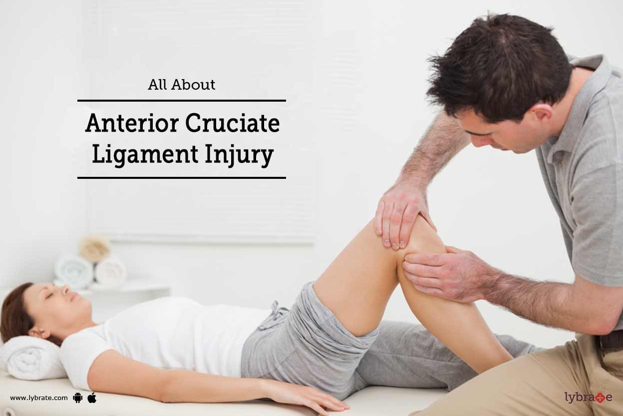
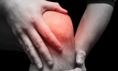
In general, anterior cruciate ligament injuries, or ACL injuries, are understood to be tears in any of the several knee ligaments joining the upper leg bone and the lower leg bone. This can vary from minor injuries, such as small ligament tears, to more serious cases, like complete tears or when the ligament and one of the bones gets displaced from the other. These injuries usually occur during sports activities, like soccer, basketball, football, gymnastics, tennis, volleyball, etc. An untreate...more
Last Updated: 6 years ago• Featured Tip
Share
Bookmark
Report

MBBS, DNB - Orthopedics/Orthopedic Surge...read more
Orthopedic Doctor•Bhubaneswar

There are many situations under which people may go through joint or muscle damage. There are other portions that may also go through the side effects of such damage. During the damage, a knee ligament might be overstretched (sprained), or at times even torn. A ligament tear can be incomplete (only a portion of the filaments that make up the tendon are torn) or complete (the tendon is torn totally). Knee tendon wounds can bring about pain, swelling, wounding and lessen the movement of your knee....more

Book appointment with top doctors for Foot Ligaments Image treatment
View fees, clinic timings and reviews

Ask a free question
Get FREE multiple opinions from Doctors
posted anonymously