Lybrate Logo
Get the App
For Doctors
Login/Sign-up
Book Appointment
Treatment
Ask a Question
Plan my Surgery
Health Feed
tab_logos
About
tab_logos
Health Feed
tab_logos
Find Doctors
tab_logos
Health Packages
Facebook Icon
Twitter Icon
Telegram Icon
Linkedin Icon

femoral artery Image Health Feed

Health Query
Share
Bookmark
Report

Hi I am 34 male my coronary angiography report. Clinical resume. Recent amwi; thrombolysed with stk here on 9.1.2017. 2d echo showed distal ivs & apex hypokinesia .mild lv systolic dysfunction. Lvef45% Procedure. 6f sheath and jl 3.5 and jr 3.5 catheter. Approach rt femoral artery. Findings Lmca. Normal artery Lad. Lad is type III and recanalised artery. Mid lad has 40-50% thrombus containing lesion distal lad has subtotal thrombotic occlusion Lcx. Non dominant and normal artery .om branches are normal Rca. Dominant and normal artery pda and plv are normal Renal. Right renal .normal artery. Left renal normal artery Conclusion. thrombotic disease in lad Advice. Aggressive medical management and life style modification as per separate No stents was placed only tablets r given to me. I want to know is treatment given to me ok. Now I am taking storvas trio 20 at night n met xl 25 in morning.

Profile Image

MBBS, MD - Internal Medicine, DM - Cardi...read more

Cardiologist•Delhi
banner-image
As per your mentioned report, you had an heart attack which seems to have been managed well due to timely thrombolysis and as per this report only minimal blockage is present in this report. Based on this report the medical advice that has been given to you is fine. But we cannot comment with 100% surety without looking at the angiography CD, because sometimes the study is either not interpreted or done correctly, but assuming that this is the report your present treatment is ok.
Last Updated: 7 years ago • Featured Quiz
Share
Bookmark
Report
Profile Image

MBBS, MS - General Surgery

General Surgeon•Kolkata
banner-image
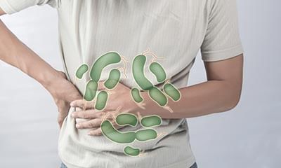
Chronic coughing can cause femoral hernia? True or False? Take this quiz to find out.
2315 people found this helpful
Asked for male, 21 years old from Kishanganj
Share
Bookmark
Report

This is my mri report please give me your precious advice about my mri reporMRI LEFT KNEE. imaging of the LEFT KNEE was performed using spin- echo, gradient echo and fast spin echo pulse sequences and high resolution T1W, FS PD and T2W were obtained in the sagittal, axial and coronal planes with STIR sequence in the coronal plane. FINDINGS: There is disruption in the continuity of fibres of anterior cruciate ligament near its femoral attachment with near horizontal orientation of the residual fibres. PDFS hyperintensity noted in the intercondylar notch. There is associated mild anterior subluxation of tibia relative to the femur. The posterior cruciate ligament is buckled otherwise normal in morphology, attachments and signal intensity. Medial and lateral collateral ligaments are intact and show normal signal intensity. Anterior horns, posterior horns and bodies of both medial and lateral menisci are normal. There is no significant collection in the knee joint. The lower end of femur, upper ends of tibial and fibula and the patella show normal signal characteristics with no evidence of any bone contusion/fracture. The muscles, tendons and fascial planes around the knee joint are normal. OPINION: Mr. FINDINGS REVEALS: Complete tear of anterior cruciate ligament near its femoral attachment.

Profile Image

MS - Orthopaedics, MBBS

Orthopedist•Pune
banner-image
Hello lybrate user,
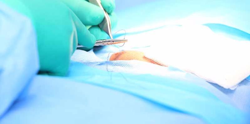
you will need a complete anterior cruciate ligament repair / reconstruction by arthroscopic techniques after you have regained normal knee joint movements.
This is a special surgery so consult a specialist for details. Follow up.
God bless you.
324 people found this helpful
Asked for male, 20 years old from new delhi
Share
Bookmark
Report

I had a knee injury and my mri result were as follow pls help examine it I am not aware of all terms and measures mri left kneestudy protocolmr imaging of the left knee was performed on a magnetom verio 3 tesla scanner using a dedicated 32-channel phased-array surface coil. Multiple sequences were obtained. Findings:cruciate ligaments :-near full thickness tear of the mid and proximal 1/3rd of acl is noted. Sprain of femoral attachment of pcl is noted. No evidence of tear. Collateral ligamentsgrade I injury / sprain of the proximal 1/3rd of lcl is noted. Mcl appears normal. Meniscus:-medial and lateral menisci appear normal in configuration and signal intensity. No evidence of tear noted. .muscles:-popliteal muscle and tendon appear normal. The quadriceps tendon and ligamentum patellae appear normal. The hoffa`s fat pad appears normal. Osseous structure:-bone marrow edema is noted in lateral and medial femoral condyle, lateral tibial condyle and fibular head. Mild joint effusion is also noted. Patella appears normal in position. Joints:-no evidence of osteoarthritis changes. Tibiofemoral and patella-femoral joints appear normal with intact articular cartilage. No obvious intraarticular loose bodies are seen. Mild joint effusion is also noted. Mri left kneeimpression: near full thickness tear of mid and proximal 1/3rd of acl. strain of femoral attachment of pcl. grade I injury of proximal 1/3rd of lcl. bone marrow edema/contusions in femur and tibia and fibular head. mild joint effusion.

Profile Image

D.P.T, BPTh/BPT, MD Acupuncture, Advance...read more

Physiotherapist•Hyderabad
banner-image
There is grade 1 injury in the knee joint, through medicines and electrotherapy it can be healed with proprr rest.
Consult ortho doc for medicines.
9 people found this helpful
Health Query
Share
Bookmark
Report
Profile Image

Fellowship in Shoulder and Knee Surgerie...read more

Orthopedist•Nagpur
banner-image
It looks like you have ignored the problem for long time. 10 years is too long period. You are just 28 and all findings of MRI are suggestive of damage to meniscus, and Knee joint which is usually seen after 50 years of age. You need to undergo Arthroscopy surgery. Meniscus should be repaired if it is repairable, though chances of repair are low as it is very old injury as per your history. Need to see MRI images to comment more.
Asked for male, 28 years old from Nashik
Share
Bookmark
Report

Right hip: there is an irregular t2 and t1 hypointense lesion in the right femoral head subarticular region in the superior articular surface. The articular surface itself is maintained. Head and neck of femur are normal and show normal signal changes, there is no marrow edema. Acetabular margins are normal. Acetabular labrum is normal. Muscles surrounding the hip joint are normal. Psoas muscles also appear normal. Greater and lesser tronchanters are normal. Hip joint space is normal. There is mild joint effusion. Sacro-iliac joints are normal. No evidence of abnormal signal changes. Left hip: there is subcortical collapse in the weight bearing portion of the left femoral head - it involves nearly 3/4th of the articular surface. There is minimal poorly defined marrow edema in the underlying bone. There is moderate effusion. Acetabular margins are normal. Acetabular labrum is normal. Muscles surrounding the hip joint are normal. Psoas muscles also appear normal. Greater and lesser tronchanters are normal. Impression: x findings suggest bilateral femoral head avn: o rt side - the lesion in small and involves about 1/3rd of the articular head, the surface is maintained. Ficat stage ii. O left side - the lesion involves 3/4th of the surface, there is subchondral collapse. Ficat stage iii.

Profile Image

MBBS (Bachelor of Medicine and Bachelor ...read more

General Physician•Delhi
banner-image
Hi the radiology report that you have uploaded has no significance without the images.
You can message me privately with the radiology reports for better help if the patient.
91 people found this helpful
book_appt_icon
Book appointment with top doctors for femoral artery Image treatment
View fees, clinic timings and reviews
chat_icon

Ask a free question

Get FREE multiple opinions from Doctors

posted anonymously
Pristyn Care Banner