Lybrate Logo
Get the App
For Doctors
Login/Sign-up
Book Appointment
Treatment
Ask a Question
Plan my Surgery
Health Feed
tab_logos
About
tab_logos
Health Feed
tab_logos
Find Doctors
tab_logos
Health Packages
Facebook Icon
Twitter Icon
Telegram Icon
Linkedin Icon

Cavernous Hemangioma Health Feed

Asked for male, 48 years old from Secunderabad
Share
Bookmark
Report

Sir, recently I did a ct scan abdomen. The details are as follows TECHNIQUE: SERIAL AXIAL SECTIONS OF WHOLE ABDOMEN 08 mm DONE WITH & WITHOUT I.V. CONTRAST FINDINGS: Liver: Normal in size and mild decreased attenuation. Two small calcific lesions 5 mm and 3 mm in subsegment 7 of liver. Small isodense area in subsegment 8, on arterial phase showing intense enhancement measuring 8 mm homogenous mild enhancement in delayed phases - Likely Hemangioma No evidence of intrahepatic biliary dilation. Portal vein normal in caliber. CBD normal in caliber. Gall Bladder: Well distended. Few small enhancing iso dense areas over fungal region of Gall bladder largest 4 mm -?Polyp? Cause. Wall thickness normal. Pancreas: Normal in size, and attenuation. No evidence of focal or diffuse lesions. Pancreatic duct normal. No evidence of peripancreatic collection and calcifications. Spleen: Normal in size and attenuation. No evidence of mass lesion. Both adrenal glands are normal in size and attenuation. Right Kidney: Normal in size and position. Few non enhancing hypodense areas largest measuring 6 mm - S/o Cysts. Few small calculi seen in mid pole largest 5 mm Pelvicalyceal system is normal. Right ureter is normal in course and caliber. Left Kidney: Normal in size and position. Few Cysts largest measuring 12 mm - S/o Cysts Small calculus of 3 mm in mid pole. Pelvicalyceal system is normal. Left ureter is normal in course and caliber. Urinary Bladder: Well distended. No evidence of filling defects. Wall thickened -?Cystitis No evidence of calculi. Prostate: Upper limit of normal in size. Small calcifications noted No free fluid in the peritoneal cavity. Bowel loops appear normal. Aorta / IVC are normal in course and caliber. Appendix visualised appear normal in caliber Spine - Small osteophytes seen, Spondylolysis of L5 vertebra, minimal disc space narrowing between L5 - S1 - Early changes of degeneration IMPRESSION: C.T. IMAGE MORPHOLOGY IS IN FAVOUR OF * Mild fatty infiltration of liver with two calcific lesions and likely small hemangioma * Few small enhancing iso dense areas over fungal region of Gall bladder largest 4 mm -?Polyp? Cause. * Bilateral small renal cortical cysts and small renal calculi * Urinary bladder wall thickened -?Cystitis. Please advise a treatment.

Profile Image

Bachelor of Ayurveda, Medicine and Surge...read more

Ayurveda•Davanagere
banner-image
Dear lybrate-user.
For your cystities fatty liver. Galblader polyp. No need to go for any surgery ayurvedic internal medicine is enough to cure completely by taking hrudaya vati (ujwala pharam) anarsha kshara (ujwala pharam), & balaguduchyadi kashaya. For 3 months. For renal calculi renal renal cyst
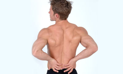
nirinjil shatavari paniyam these medicine will cure your problem cures completely. For your degenerative changes in Lumbar spine need to take panchakarma once.
Asked for male, 25 years old from Bangalore
Share
Bookmark
Report
Profile Image

MBBS

General Physician•Jalgaon
banner-image
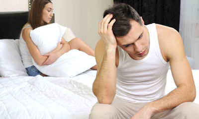
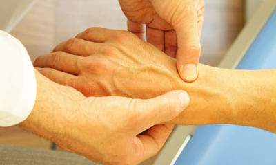
Please It is natural to take time to refill the cavernous sinuses of penis to get erected, after one episode of ejaculation Body takes its own time.
154 people found this helpful
Health Query
Share
Bookmark
Report
Profile Image

C.S.C, D.C.H, M.B.B.S

General Physician•Alappuzha
She may be having a birthmark namely hemangioma. Please send photo and it can be removed by cautery though it may leave a small scar. May be it is a brown mole. Send photo.
342 people found this helpful
Asked for male, 3 years old from Khagaria
Share
Bookmark
Report
Profile Image

MD - Paediatrics, MBBS

Pediatrician•Jaipur
banner-image
This could be congenital hemangioma (dilated blood vessels below skin)
this may bleed a lot if injured so take utmost care to prevent trauma.
1000 people found this helpful
Health Query
Share
Bookmark
Report
Profile Image

MBBS, MS - General Surgery, FIAGES(Fello...read more

General Surgeon•Ghaziabad
banner-image
Hello. It can be a small hemangioma on the glans. please consult any surgeon nearby place. Don't ignore.
149 people found this helpful
Health Query
Share
Bookmark
Report
Profile Image

C.S.C, D.C.H, M.B.B.S

Cardiologist•Alappuzha
Hemangioma usually heals itself or will stay but is harmless. If you want to remove it you need laser treatment
Asked for female, 45 years old from Warangal
Share
Bookmark
Report

I am 45 years old (female. To of my knowledge I was fell down due to slippage of my foot six to seven times in past two years and I was neglected it .But since last six months I had severe pain in my left leg from knee to below and went to local physician doctor at warangal but of vain. On advice of him I was went to local neuro physician and MRI scan was done .The finding of MRI scan are reproduced as it is for your advise and providing medical treatment to me. 1) Degenerative changes are seen in the form of marginal osteophytes and partial disc desiccation at multiple levels. 2) Well defined T1 & T2 hypertensive lesion seen in L3 vertebral body- likely hemangioma. 3) Modic type II end plate changes are seen in L4 vertebra. 4) T2 & STIR hyperintense and T1 hypointense signal seen in anteroinferior aspect of L4 vertebral body. 5) Diffuse annular disc bulges noted at L1-L2, L2-L3, &L3-L4 levels causing indentation of thecal sac. 6) Diffuse asymmetric annular disc bulges with posterior disc protrusion is noticed at L4-L5 level causing indentation thecal sac, narrowing of bilateral neural foramen (left>right) with impingement of bilateral traversing nerve roots. 7) Mild annular disc bulges noted at L5-S1 level causing indentation of thescals sac. Thanking you sir and hoping the early advice.

Profile Image

MPT, BPT

Physiotherapist•Noida
banner-image
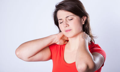
Quadriceps Exercises- Lie straight, make a towel role and put it under the knee, press the keen against the role, hold it for 20 secs. Repeat 20 times twice a day. This will help relieve some pain.
Core Strengthening Exercise- Straight Leg Raised With Toes Turned Outward, repeat 10 times, twice a day.
Hams Stretching- lie straight, take the leg up, pull the feet towards yourself, with a elastic tube or normal belt. repeat 10 times, twice a day.
57 people found this helpful
Asked for male, 35 years old from Coimbatore
Share
Bookmark
Report
Profile Image

Erasmus Mundus Master in Adapted Physica...read more

Physiotherapist•Chennai
banner-image
What is a hemangioma? Spinal hemangiomas are benign tumors that are most commonly seen in the mid-back (thoracic) and lower back (lumbar. At upmc, we treat hemangiomas with surgical removal (resection) of the tumor or the affected vertebra, and radiation therapy to treat pain. Treatment for hemangiomas depends on the size and location of the tumor. At upmc, we use a combination of stopping blood flow to the tumor (embolization), surgical removal of the tumor, and radiation therapy.
Radiation...more
Asked for Male, 22 years old from Bangalore
Share
Bookmark
Report

BHMS

Sexologist•Noida
banner-image
Masturbation every day very harmful muscles of penis affected 1 corpus cavernous 2 vascular muscles 3 corpus spongiosum Avoid to masturbation 1 impotency.
83 people found this helpful
Health Query
Share
Bookmark
Report

Hi, I have been having numbness in my right great toe for the last 2-3 months. In addition, I do have back stiffness at times and pain especially when I exert my back, let's say during long drives or any activity that impacts my lower back. In the last 1 month, I was into yoga with the help of an instructor and not sure if I really did it wrong. I have the habit of doing Vajrasana esp post meals and even sit in ardha padmasana when possible. But in the last 1 month, have developed pain in both my legs esp when I sit cross legged. It's not a lingering pain though but radiates when I release my leg from a cross legged position. Pain is higher in the left leg. Thanks to physio and I feel better. My physio suggested a MRI LS spine. Impressions read as follows: FINDINGS: There is loss of lumbar lordosis. Small anterior and lateral marginal osteophytes are seen from L1 to L5 vertebral level. T1W and TZW hyperintense hemangioma ls noted In L1 and L5 vertebral bodies. At L2-L3 level: diffuse disc bulge indenting thecal sac narrowing bilateral neural foramina without any nerve root compression. At L3-L4 level: diffuse disc bulge indenting thecal sac narrowing bilateral neural foramina abutting left exiting nerve roots. Ligamentum flavum hypertrophy is noted at this level. At L4-L5 level: central and bilateral paracentral disc bulge indenting thecal sac narrowing bilateral neural foramina abutting bilateral exiting nerve roots. Ligamentum flavum hypertrophy ls noted at this level. At L5-S1 level: central and bilateral paracentral disc bulge indenting thecal sac narrowing bilateral neural foramina abutting bilateral exiting nerve roots. Rest of the lumbosacral vertebral bodies show normal marrow signal. The facet joints and posterior elements are normal. Rest of the intervertebral discs are normal in signal intensity. The spinal cord is normal in course, caliber and signal intensity and ends in L1, conus medullaris is normal. I would request advice on the following: 1.kindly Explain this condition in simple terms 2. Suggested course of treatment 3. Implications of this condition if it continues to persist 4. Do s and don't s Thanks.

Profile Image

Bachelor of Unani Medicine and Surgery (...read more

Ayurveda•Kanpur
banner-image
ligaments and nerves ka issue hai ( in simple language )

ras raj ras 125 mg twice a day
vatari avleh 10 gm twice a day
relief in 3-4 days and for complete cure take it for 60 days only
avoid oily and spicy food
book_appt_icon
Book appointment with top doctors for Cavernous Hemangioma treatment
View fees, clinic timings and reviews
chat_icon

Ask a free question

Get FREE multiple opinions from Doctors

posted anonymously
Pristyn Care Banner