Lybrate Logo
Get the App
For Doctors
Login/Sign-up
Book Appointment
Treatment
Ask a Question
Plan my Surgery
Health Feed
tab_logos
About
tab_logos
Health Feed
tab_logos
Find Doctors
tab_logos
Health Packages
Facebook Icon
Twitter Icon
Telegram Icon
Linkedin Icon

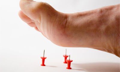
Bone marrow Image Health Feed

Asked for female, 24 years old from Chittoor
Share
Bookmark
Report
Profile Image

MD - Homeopathy, BHMS

Homeopath•Vadodara
banner-image
Cleanser should be choosen as per skin type and not with the open pores size. There are 4 type of skin. Dry, oily, combination and normal. So chhoose as per your skin type. You can consult me at Lybrate for homoeopathic treatment and further guidance about skin care.
22 people found this helpful
Asked for female, 67 years old from Panchkula
Share
Bookmark
Report
Profile Image

MBBS

General Physician•Jalgaon
banner-image
Please send me ultrasound reports meanwhile take plenty of water daily take sy neeri by aimil pharma 20 ml in 200 ml water three times a day for 3 weeks.
27 people found this helpful
Asked for female, 42 years old from Hisar
Share
Bookmark
Report
Profile Image

BHMS

Homeopathy Doctor•Noida
banner-image
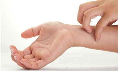
It can be because of dermatitis/ eczema or allergy or dryness or fungal infection or psoaiasis etc. I need details of case n preferably pics of affected area. In the meanwhile follow this
moisturize frequently.
Avoid sudden changes in temperature or humidity.
Avoid sweating or overheating.
Reduce stress.
Avoid scratchy materials, such as wool directly in contact with skin.
Avoid harsh soaps, detergents, and solvents.
Be aware of any foods that may cause an outbreak an...more
14 people found this helpful
Asked for male, 20 years old from new delhi
Share
Bookmark
Report

I had a knee injury and my mri result were as follow pls help examine it I am not aware of all terms and measures mri left kneestudy protocolmr imaging of the left knee was performed on a magnetom verio 3 tesla scanner using a dedicated 32-channel phased-array surface coil. Multiple sequences were obtained. Findings:cruciate ligaments :-near full thickness tear of the mid and proximal 1/3rd of acl is noted. Sprain of femoral attachment of pcl is noted. No evidence of tear. Collateral ligamentsgrade I injury / sprain of the proximal 1/3rd of lcl is noted. Mcl appears normal. Meniscus:-medial and lateral menisci appear normal in configuration and signal intensity. No evidence of tear noted. .muscles:-popliteal muscle and tendon appear normal. The quadriceps tendon and ligamentum patellae appear normal. The hoffa`s fat pad appears normal. Osseous structure:-bone marrow edema is noted in lateral and medial femoral condyle, lateral tibial condyle and fibular head. Mild joint effusion is also noted. Patella appears normal in position. Joints:-no evidence of osteoarthritis changes. Tibiofemoral and patella-femoral joints appear normal with intact articular cartilage. No obvious intraarticular loose bodies are seen. Mild joint effusion is also noted. Mri left kneeimpression: near full thickness tear of mid and proximal 1/3rd of acl. strain of femoral attachment of pcl. grade I injury of proximal 1/3rd of lcl. bone marrow edema/contusions in femur and tibia and fibular head. mild joint effusion.

Profile Image

D.P.T, BPTh/BPT, MD Acupuncture, Advance...read more

Physiotherapist•Hyderabad
banner-image
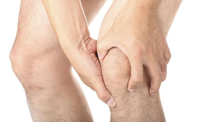
There is grade 1 injury in the knee joint, through medicines and electrotherapy it can be healed with proprr rest.
Consult ortho doc for medicines.
9 people found this helpful
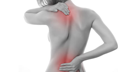
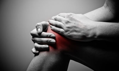
Asked for male, 62 years old from Ahmedabad
Share
Bookmark
Report

Oblique vertical fracture with marrow oedema are seen involving the anteroinferior aspect of the glenoid articular surface, extending from 2: 00 to 6: 00 o'clock position. The fragment measures 30 x 12 x 10 mm in si x ap x tr dimensions. Depression is seen along the articular surface with signal abnormality within the overlying cartilage suggesting chandra injury. Minimal anterior and anteroinferior displacement of the fracture fragment is seen relative to the rest of the glenoid. Linear loose fracture fragment noted along the posterior inferior aspect of the humeral head. There is a complete tear of the anterosuperior labrum, from approximately 1: 00 to 3: 00 o'clock position. Ill-defined hyperintense signal is seen on the pd images at the base of the anterior aspect of the superior labrum. The anteroinferior glenoid labrum is intact and attached to the fractured fragment. Sprain injury involving the superior and middle glenohumeral ligaments. Hill-sachs lesion measuring approximately 18 x 9 mm[ tr x si ] and 4 mm in depth is noted associated with marrow contusion. Moderate joint effusion. Intact rotator cuff tendons, muscle bellies, superior labrum and lhbt.

Profile Image

MBBS, Basic Life Support (B.L.S), Advanc...read more

General Physician•Delhi
The main subcortical limbic brain regions implicated in depression are the amygdala, hippocampus, and the dorsomedial thalamus. Both structural and functional abnormalities in these areas have been found in depression. But we have to look into the details of the problem. Let's have a detailed discussion to Ensure proper treatment.
19 people found this helpful
Asked for male, 25 years old from Kangra
Share
Bookmark
Report

I am 25 years old. I have strong pain in left knee joint. I consulted a doctor .he told me for MRI. But not only told me for physiotherapy .but I am doing from last 1 month but there is no effect. Please suggest me something after watching my MRI report- MRI LEFT KNEE JOINT STUDY PROTOCOL : SAGITTAL : PD, GRE // CORONAL : STIR, T1W // AXIAL :T2 TSE FINDINGS : There is mild effusion in knee joint extending into suprapatellar and prepatellar recess. Femorotibial articular surfaces and joint space are normal. Visualised bones are normal in cortical outline and marrow signal intensity. Grade II signal change on PD images reaching upto capsular surface is seen in the posterior horn of medial meniscus. Lateral meniscus is normal in outline and signal intensity. Anterior and posterior cruciate ligaments are normal in course with normal parenchymal signal intensity. Quadriceps femoris tendon and patellar tendon are normal with normal signal intensity. Patella is normal in location. Patellar articular cartilage is normal in thickness, outline and signal intensity. Medial and lateral collateral ligaments are normal with no evidence of tear. Peri-articular and para-articular muscles reveal normal parenchymal signal intensity and well-preserved intermuscular fat planes. IMPRESSION: FINDINGS ARE SUGGESTIVE OF *GRADE II SIGNAL CHANGE IN POSTERIOR HORN OF MEDIAL MENISCUS. *MILD KNEE JOINT EFFUSION. PLEASE CORRELATE CLINICALLY.

Profile Image

BPTh/BPT

Physiotherapist•Ghaziabad
banner-image
Hello according to your report your posterior meniscus is tear. You have to need knee braces to maintain knee in extension.
Knee braces sote time v laga k hi sona.
While walking donot give full weight.
In physiotherapy session you take ultrasonic therapy for 12 mints on posteriorly of knee.
Ift therapy for 15 mints posteriorly knee.
In home you take ice or cold pack for 20 mints on back of knee.
Take rest for 4 weeks.
Because ligaments tear take 6-8 of healing time so...more
Asked for Male, 53 years old from Moga
Share
Bookmark
Report

Mri result cervical issues i’m 26 male. I was having little numbness in my right should and leg. Got nerve test and mri. Nerve test was normal. Even doctor said the mri is normal. But when I saw the report at c3/c4 it shows something. Can you guys help. Is it normal mri mri says: technique: sagittal t1, sagittal t2, sagittal t2 repeat, axial t2 and axial t2 3d merge. Findings: despite repeat sequences image quality is moderately degraded by motion artifact. Alignment is stable. Vertebral body heights are preserved. No suspicious bone marrow signal changes are present. The visualized posterior fossa structures are unremarkable. The visualized soft tissues of the neck demonstrate no focal abnormality. Normal bilateral vertebral artery flow voids are demonstrated. The axial t2 sequences are significantly degraded by motion which reduces sensitivity and specificity of this examination for characterizing spinal or neural foramina narrowing. C2/c3: unremarkable. C3/c4: small disc osteophyte complex contributes to no significant spinal or neural foramina narrowing. C4/c5: unremarkable. C5/c6: unremarkable. C6-c7: unremarkable. C7/t1: unremarkable. Impression: limited examination due to motion. No large disc osteophyte complex present. No definite spinal stenosis or cord compression is present. Intrinsic signal abnormality within the cord is difficult to exclude on this examination due to motion artifact. No major neural foramina stenosis is present. Mild neural foramina stenosis can not be definitively excluded due to motion artifact on the critical axial sequences.

Profile Image

MBBS , MD - Biochemistry

General Physician•Lucknow
banner-image
The osteophyte complex at c3/c4 may or may not be the reason for your symptoms. In any case your symptoms should go away with rest in a 2-3 weeks. However if the symptoms persist or worsen you should again consult a neurologist (dm neurology)
23 people found this helpful
Health Query
Share
Bookmark
Report
Profile Image

MD - Homeopathy, BHMS

Homeopathy Doctor•
banner-image
Doctors have no use of extra bone marrow taken... They don't get any benefit by getting more... So don't listen to stupidity of others...
Yes not all doctors are good.. but they are not killers...
book_appt_icon
Book appointment with top doctors for Bone marrow Image treatment
View fees, clinic timings and reviews
chat_icon

Ask a free question

Get FREE multiple opinions from Doctors

posted anonymously
Pristyn Care Banner