Get the App
For Doctors
Login/Sign-up

About
Health Feed
Find Doctors
Health Packages

Bone marrow Image Questions
Health Query
Share
Bookmark
Report
You should take acupressure treatment and take biochemic Nat phos200x + kali Mur 200x 4 tab each thrice a day with warm water and take it 5 day's and consult private online.
258 people found this helpful
Asked for male, 20 years old from new delhi
Share
Bookmark
Report

There is grade 1 injury in the knee joint, through medicines and electrotherapy it can be healed with proprr rest.
Consult ortho doc for medicines.
Consult ortho doc for medicines.
9 people found this helpful
Health Query
Share
Bookmark
Report
Its always suggessted to do only cold pack as it helps to reduce the inflammation. Hot pack increases the swelling.
47 people found this helpful
Asked for male, 62 years old from Ahmedabad
Share
Bookmark
Report

The main subcortical limbic brain regions implicated in depression are the amygdala, hippocampus, and the dorsomedial thalamus. Both structural and functional abnormalities in these areas have been found in depression. But we have to look into the details of the problem. Let's have a detailed discussion to Ensure proper treatment.
19 people found this helpful
Asked for male, 25 years old from Kangra
Share
Bookmark
Report
Hello according to your report your posterior meniscus is tear. You have to need knee braces to maintain knee in extension.
Knee braces sote time v laga k hi sona.
While walking donot give full weight.
In physiotherapy session you take ultrasonic therapy for 12 mints on posteriorly of knee.
Ift therapy for 15 mints posteriorly knee.
In home you take ice or cold pack for 20 mints on back of knee.
Take rest for 4 weeks.
Because ligaments tear take 6-8 of healing time so...more
Asked for Male, 53 years old from Moga
Share
Bookmark
Report
The osteophyte complex at c3/c4 may or may not be the reason for your symptoms. In any case your symptoms should go away with rest in a 2-3 weeks. However if the symptoms persist or worsen you should again consult a neurologist (dm neurology)
23 people found this helpful
Health Query
Share
Bookmark
Report
Doctors have no use of extra bone marrow taken... They don't get any benefit by getting more... So don't listen to stupidity of others...
Yes not all doctors are good.. but they are not killers...
Yes not all doctors are good.. but they are not killers...
Asked for male, 21 years old from Tirupati
Share
Bookmark
Report
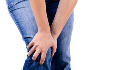
Man you r injured a lot start physiotherapy immediately you can go to college with hinge knee brace on knee
stop cross sitting avoid sitting on ground.
stop cross sitting avoid sitting on ground.
Asked for male, 21 years old from Jamshedpur
Share
Bookmark
Report

1.Masturbation is a normal way of relieving sexual tension.
2.It is, not harmful if done in moderation, preferably 2-3 times a week.
3.Excess indulgence may cause physical problem like weakness, tiredness, loss of memory, lack of energy, lack of concentration, loss of facial glow and sexual problem like erectile dysfunction,premature ejaculation,testicular pain, decrease sperm count.etc
Masturbation does not cause bone marrow depression
3036 people found this helpful
Asked for male, 21 years old from Delhi
Share
Bookmark
Report
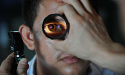
Hello. There is no medication or food for improving eye sight or to get rid of glasses.
Get your vision tested and wear glasses.
If not interested in glasses then you can try
a. Contact lens
b. Lasik laser
as your age is now 19 years so your number will not increase now whether you wear glasses or not as number becomes stable at age of 18-20 yrs.
Having said that I advise you to eat balanced diet including green vegetables and seasonal fruits for keeping eyes and body hea...more

Book appointment with top doctors for Bone marrow Image treatment
View fees, clinic timings and reviews

Ask a free question
Get FREE multiple opinions from Doctors
posted anonymously