Get the App
For Doctors
Login/Sign-up

About
Health Feed
Find Doctors
Health Packages

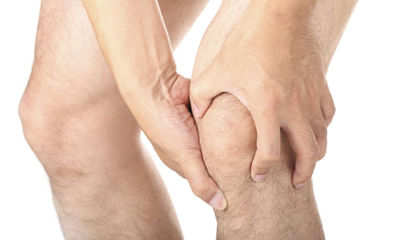
Achilles Tendon Image Questions
Asked for male, 26 years old from Lucknow
Share
Bookmark
Report

Prevention
•wear shoes that fit properly and support the foot.
•wear the right shoes for physical activity.
•stretch your muscles before exercising.
•pace yourself during physical activity.
•maintain a healthy diet.
•rest when you feel tired or when your muscles ache.
•maintain a healthy weight.
Treatment
•rest as much as possible.
•apply ice to the heel for 10 to 15 minutes twice a day.
•take over-the-counter pain medications.
•wear shoes that fit...more
98 people found this helpful
Health Query
Share
Bookmark
Report
Physical therapy. A physical therapist can instruct you in a series of exercises to stretch the plantar fascia and achilles tendon and to strengthen lower leg muscles, which stabilize your ankle and heel. A therapist might also teach you to apply athletic taping to support the bottom of your foot. Apply ice over the area.
13 people found this helpful
Asked for male, 25 years old from Kangra
Share
Bookmark
Report
Hello according to your report your posterior meniscus is tear. You have to need knee braces to maintain knee in extension.
Knee braces sote time v laga k hi sona.
While walking donot give full weight.
In physiotherapy session you take ultrasonic therapy for 12 mints on posteriorly of knee.
Ift therapy for 15 mints posteriorly knee.
In home you take ice or cold pack for 20 mints on back of knee.
Take rest for 4 weeks.
Because ligaments tear take 6-8 of healing time so...more
Asked for male, 20 years old from new delhi
Share
Bookmark
Report

There is grade 1 injury in the knee joint, through medicines and electrotherapy it can be healed with proprr rest.
Consult ortho doc for medicines.
Consult ortho doc for medicines.
9 people found this helpful
Asked for male, 32 years old from Allahabad
Share
Bookmark
Report

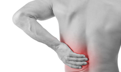
Wrist pain is often caused by sprains or fractures from sudden injuries. But wrist pain can also result from long-term problems, such as repetitive stress, arthritis and carpal tunnel syndrome.
Because so many factors can lead to wrist pain, diagnosing the exact cause can be difficult, but an accurate diagnosis is essential for proper treatment and healing.
Imaging tests such as x-ray, mri, ct scan or ultrasounds can be suggested to identify the root cause.
If imaging test results ar...more
24 people found this helpful
Health Query
Share
Bookmark
Report
CSR is also known as Macular oedema due to unknown cause but some factor are responsible i.e Stress DM etc if not cure within time it cause permanent damage in macular region and also chances of recurrence in alopathy inj avastin is only options, in ayurveda their is much better options available. through ayurvedic treatment you can improve your vision and avoid recur ency also avoid from much costly injection
some suggestions take healthy diet, avoid spicy foods, avoid preservative contai...more
some suggestions take healthy diet, avoid spicy foods, avoid preservative contai...more
374 people found this helpful
Asked for male, 26 years old from Bangalore
Share
Bookmark
Report
Well start doing yoga, do regular exercise, do meditation, you need proper homeopathic treatment for this purpose along with counseling.
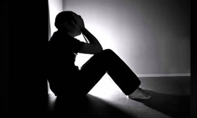
Asked for male, 35 years old from Patna
Share
Bookmark
Report
Hello,
If the images are repetitive, not under your control, causing distress so much that its hampering your day to day life than you might have OCD. The behavior you are repeating to get rid of these images are known as compulsive acts. OCD can affect anyone and it can be treated with medication and behavior therapy.
Whenever these thoughts come try involving yourself in some other activity or start repeating in your mind 3 times go away. It might help. Contact nearby neuropsychiatric ...more
Asked for male, 28 years old from Noida
Share
Bookmark
Report
its your thinking, you need to change your thinking. change your mind about sex, its normal for all but yur behavior is not normal. thanks
Health Query
Share
Bookmark
Report

This is a psychological condition not much worry about it,,do yoga,, meditation,make urself bzy in ur work,, don't think much,,take it lightly,,with this u need proper homoeopathic treatment to cure ur problem permanently

Book appointment with top doctors for Achilles Tendon Image treatment
View fees, clinic timings and reviews

Ask a free question
Get FREE multiple opinions from Doctors
posted anonymously