
Get the App
For Doctors
Login/Sign-up

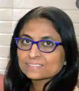
Dr. Usha Seth
Radiologist39 Years Exp.
MBBS, MD - Radio Diagnosis/Radiology
Delhi
₹ 0 at clinic
Personal Statement
I believe in health care that is based on a personal commitment to meet patient needs with compassion and care...read more
Doctor Information
Speciality
- Radiologist
Other treatment areas
- Radiologist
Education
- MBBS , Rehman Medical College , 1986
- MD - Radio Diagnosis/Radiology , Rehman Medical College , 1991
Languages spoken
- English
- Hindi
Clinic Location
E-56, Ground Floor, Narayana Vihar, Delhi, Delhi
Clinic of Dr. Usha Seth
| Clinic's Name | Fees |
|---|---|
| Dr. Seth Child & Ultra Sound Clinic | - |
Get Help
Services