
Diagnosis Of Spine-Related Problems


Hi,
I am Dr. Sidharth Verma. I am a pain physician and I treat patients who are suffering from pain. Often patients ask me what is their diagnosis and I tell them and then they are confused because what I tell them is like you have C3, C4 disc problem and then we need to do this or that and they get very confused, so I thought I will make a video so that they can understand and all of you can understand what is this terminology.
So our spine is made up of small bones which is known as vertebrae. These bones they join together at various joints and then they make the whole spine. This spine is a very important organ because it is used in whether you are lying down, whether you are standing, whether you are sitting all the time you are using your spine and without your spine you cannot even stand, you see those people who have issues with spine they cannot even stand or cannot even work so it is a very vital organ in the body and traditionally it has also been emphasized on the health of the spine.
Now let's move to the constituents, the spine is mainly composed of the bone which are this vertebra, it can be divided into four major regions one is the cervical that is a spine of the neck. So the bones in the neck they are known as spine, the cervical spine. Now let's move a little below the bones of the thorax are known as the thoracic spine. Thorax means the area below from your neck to your lower back that is the area of your chest and upper back, so these bones are named as thoracic spine. Now let's move a little down this portion is known as the lumbar spine and I have explained about the lumbar spine in my other video which you can see on my lybrate website.
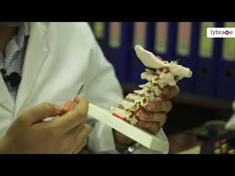
Now coming back to the cervical spine, if you see in the cervical spine and they are small bones which are joined together at three joints each and they are enclosing the spinal cord in between at various holes which is known as the foramen, the spinal nerves they come out and supply the various parts of the body. So if you see there are various pain generators which can be there in these bones but let us first label these bones.
So this model is a model of the cervical spine that is the bones inside the neck. So these are the bones and they are labelled as or named as cervical one or the C1, C2, C3, C4, C5, C6 and C7 so these 7 bones makeup the cervical spine and if you see the first one is very different than the rest of the other bones and that's why it is very atypical type of a vertebrate that is it is not like the others for all practical purposes it is labelled as C1, C2, C3, C4, C5, C6, C7. So when you see a report which mentions there is a C2-3 disc prolapse that means this is a second and third and this is the disc in between and that now the height has decreased and it has come off or it has been damaged and then it is pressing on the nerves.
So this is the disc in between the 3rd and 4th so C3-4, this C5-6 so this is a disc in between the C5 and C6. If you these vertebrae they also have small holes through which these nerves are coming out. so, this nerves are coming out of these small holes, these holes are known as the foramen or the intervertebral foramen.
So, there is another structure which is this artery which is going to the brain, so it takes the blood supply from the heart to the brain. It also passes very close, see how close these nerves are to this artery. So, it is very close and when your pain physician will explain to you some procedures they will tell you the risk involved with these procedures because this arteries very close by. So now if you see on the posterior portion there are two joints which is the right and the left facet joint and on the anterior there is one joint which is made up of this intervertebral discs. So, again like in lumbar, cervical also have 3 joints in between the two bones. So this front joint this position is known as the intervertebral disc and the posterior joints are the facet joint. This is a right facet joint, this is the left facet joint in between C2 and C3 cervical vertebra, this is the right facet joint and the left it is in between C3 and C4 cervical vertebra and these are the joints between C4 and C5, C5 and C6, C6 and C7 cervical vertebra. So if you see this structure is passing, that is a spinal cord is passing inside these bones through a canal and it goes down to the rest of the body.
Now the important thing to note here is there is one structure which is not seen here in this model and that is the muscles, so if you see for example a big muscle attaches from here and also here. So it is this thick and supports the whole of the cervical spine. It results in a lot of movement at this portion but can also cause pain if the person is not exercising or over using it or under using it and that can result in neck pain. Some types of neck pain can also radiate to the head because some of the portion of the head nerve supply comes from these neck nerves only.
I have explained to you the cervical spine in an easy and simple manner and now you will be able to understand after seeing your x-ray reports, after talking to a doctor, what is this C1, C2, C3, C4, C5 and what it means? and if you have any more queries you are most welcome to contact us. If you are suffering from neck pain please feel free to contact us. If you want to know more, you can visit our lybrate website and get in touch with us.
Thank you


+1.svg)
