
Get the App
For Doctors
Login/Sign-up
Last Updated: Oct 23, 2019
BookMark
Report
Retinal Detachment - 3 Most Common Types!


Dr. Shalini ShettyOphthalmologist • 31 Years Exp.Fellowship of the Royal College of Surgeons (FRCS), MS - Ophthalmology, MBBS
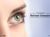
Retinal detachment is an emergency eye condition in which the retina at the back of the eye gets separated from the surrounding tissue and pulls away from its normal position. The retina acts as a light-sensitive wallpaper in the eye, providing a lining for the inside of the eye wall and sending visual signals to the brain. As the retina can't work properly under these conditions, one can permanently lose vision if the detached retina is not repaired immediately.
- During the retinal detachment, the retinal cells gets separated from the layer of blood vessels which provides oxygen and nourishment. Usually, it begins in form of small torn area of retina known as retinal tears or retinal breaks. This condition, if not treated, leads to retinal detachment and finally permanent vision loss.
- Retinal detachment has tell-tale warning signs like an increase in sudden appearance of floaters resembling cobwebs floating in field of vision. It can be coupled with flashes of light or curtain from any direction causing a loss of vision.
- Retinal detachment is of three types. The most common form is Rhegmatogenous retinal detachment where a tear allows fluid to get under retina and prevents nourishment to reach retina from retinal pigment epithelium by separating them. In Fractional form, scar tissue on the retina's surface shrinks causing it to separate from the retinal pigment epithelium. This form is most prevalent with diabetes patients. Lastly, in case of Exudative retinal detachment, the fluid leaks into the area under retina without a tear or breaks in the retina. Retinal diseases or trauma to the eye are main causes for Exudative retinal detachment.
- Although a person of any age can suffer from retinal detachment, but it is more prevalent in people over the age of 40. People suffering from degenerative myopia or lattice degeneration are more prone to this medical condition. People with family history of retinal detachment are also likely to suffer from the same.
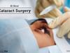
- Retinal detachment can be treated in many ways. The most common form is the Laser surgery in which small tears and hole are joined back to the retina. Another method is Cryopexy in which the area around the hole in frozen and helps reattach the retina. Both the above procedure are performed at ophthalmologist's clinic.
- Sometimes, one may have to opt for Scleral buckle in which a tiny synthetic band is attached to the outside of the eyeball which gently pushes the wall of the eye in toward the centre of the eye placing the eye wall very close to the detached retina. Another option is vitrectomy surgery to replace the vitreous that fills the centre of the eye and helps the eye maintain a round shape.
- A retinal detachment is an emergency medical condition and must be treated immediately to save one's vision. Most people have been successfully treated for retinal detachment, but ophthalmologists cannot always predict how vision will turn out. The visual outcome will not be known for up to several months after surgery. However the results are best when the retinal detachment is treated as soon as possible. If you wish to discuss about any specific problem, you can consult an Ophthalmologist.
240 people found this helpful


RELATED ISSUES
RELATED SPECIALITIES
Ask a free question
Get FREE multiple opinions from Doctors
posted anonymously


+1.svg)
