Get the App
For Doctors
Login/Sign-up

Health Query
Share
Bookmark
Report
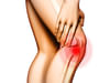
I have problem with my knees. I am in pain when walking, climbing stairs and exercising. I have done mri to both knees and the radiology report is as following, I need to consult an ortho doctor to discuss the mri both knees findings and have a solution: mri right knee (1.5t mri) history: pain in both knees (left > right) findings: there is minimal right knee joint effusion. Small area showing mild subchondral degenerative change in the articular part of the medial condyle of femur. Grade I changes in the posterior horn of the medial meniscus. The lateral meniscus is normal. Medial and lateral collateral ligaments are normal. Anterior and posterior cruciate ligaments are normal. Tibial, femoral condyle and upper end of fibula are normal. No evidence of loose bodies. posture-lateral corner structures including the popliteus tendon popliteo-fibular ligament are normal. Normal patella alignment is seen. The extensor tendons including the quadriceps and patellar tendons are normal. Proximal tibia-fibular joint is normal muscles surrounding the knee joint are normal. Impression: v there is minimal right knee joint effusion. V small area showing mild subchondral degenerative change in the articular part of the medial condyle of femur. V grade I changes in the posterior horn of the medial meniscus. The findings are of very early degenerative changes and are not a serious problem. Suggested clinical correlation mri left knee (1.5t mri) history: pain in both knees (left > right) there is minimal left knee joint effusion. There are few small subchondral cysts measuring 2 mm in the anterior part of the lateral tibial plateau area. There is mild suspicious focal hyperintensity in the upper part of the posterior cruciate ligament. The anterior cruciate ligament is normal. Medial and lateral meniscus is normal. Medial and lateral collateral ligaments are normal. Tibial, femoral condyle and upper end of fibula are normal. No evidence of loose bodies. posture-lateral corner structures including the popliteus tendon popliteo-fibular ligament are normal. Normal patella alignment is seen. The extensor tendons including the quadriceps and patellar tendon s are normal. Proximal tibia-fibular joint is normal muscles surrounding the knee joint are normal. Impression: v there is minimal left knee joint effusion. V there are few small subchondral cysts measuring 2 mm in the anterior part of the lateral tibial plateau area. V there is mild suspicious focal hyperintensity in the upper part of the posterior cruciate ligament. The findings are of early degenerative changes and are not a serious problem. Suggested clinical correlation there are very early degenerative changes in both knees (left > right). However, the findings are not likely to cause severe knee pain. Regards and thanks raha.
1Doctor Answered


91% (697 ratings)
Ask Free Question
Respected Lybrate user. From the above mentioned details (mri). Very little problem is present in your both knees but don't be neglectd. Because in near future the all above's changes are converted into your knees arthritis. Please be careful. Take treatment for your orthopaedic doctor and take advices, precautions and preventive measures. In my side you are taking precautions, preventive measures, life style modifications, dietitian/nutritionist advices. Take diet chart. Ergonomical, postural corrections are required. Which tells your orthopaedic doctor. Or physical therapist. But don't be forget and don't be neglect.
0 person found this helpful

Was this answer helpful?
YESSOMEWHATNO
Take help from the best doctors

Suggestions offered by doctors on Lybrate are of advisory nature i.e., for educational and informational purposes only. Content posted on, created for, or compiled by Lybrate is not intended or designed to replace your doctor's independent judgment about any symptom, condition, or the appropriateness or risks of a procedure or treatment for a given person.

Book appointment with top doctors for Knee Pain treatment
View fees, clinic timings and reviews
Ask a free question
Get FREE multiple opinions from Doctors
posted anonymously


Treatment Enquiry
Get treatment costs, find best hospitals/clinics and know other details