AR hospital orthopedic
Orthopaedic Clinic


About Clinic
We are dedicated to providing you with the personalized, quality health care that you deserve....read more
Clinic Timing
Clinic Location
Videos

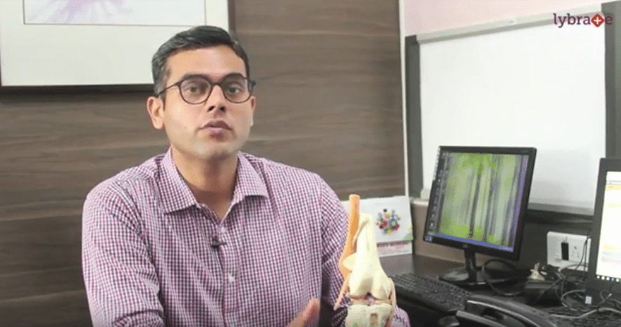
Hi!
I am Dr. Shailesh Mishra, I am orthopaedic surgeon. I am specialized in arthroscopic surgery and joint replacement. Today I am going to talk about shoulder injuries particularly shoulder dislocation. Dislocation means popping up of joint that is joint is made up of two bones together joined by the fibrous tissue. The shoulder joint is made up of glenoid; glenoid which is part of scapula bone and humeral head which is part of humerus bone. These two are held together by fibrous tissue which is capsule and the thicker portion of capsule is labrum. In case of a dislocation, these capsular-labral structures are tonged and this humeral head pops out of the joint. Why shoulder dislocation is the most common dislocation? The shoulder is made up of two bones where glenoid is smaller as compared to the humeral head, because of this mismatch in the two bones, the shoulder joint is inherently unstable joint and hence this dislocation is the most common. What are the types of dislocation? The dislocation can be anterior, posterior or inferior.
The dislocation can be partial where not the entire head has come out, only the half of head has come out and it reduces back, it can be complete dislocation where the entire humeral head pops out of the joint. Typically, patient presents with pain around the shoulder joint, swelling, deformity where the roundness of the shoulder is lost, patient is not able to touch the opposite shoulder, the rotations of the shoulders are restricted. Causes of the shoulder dislocation: shoulder dislocation can occur because of the sudden blow as it is during the vehicular accident, because of the sudden force around the shoulder joint like in sports injuries like hockey, football, rugby. It can occur also because of the sudden fall where the patient falls on the shoulder joint and because of sudden force shoulder joint pops out of the capsule. Treatment for the shoulder dislocation in case of the acute event the manual reduction of the shoulder joint is recommended. The reduction should be under sedation or general anaesthesia. Manual reduction without anaesthesia or sedation can cause more damage to the shoulder joint because of the contraction of the muscle along the shoulder joint.
When there are two or more episodes of dislocation that is recurrent dislocation of shoulder joint it requires a surgery. There is a dictum in the shoulder once dislocated always dislocated that is that means once a dislocation has occurred the capsular-labular structure which is damaged do not repair on their own and repeated dislocation can occur. There is almost 80-90% of the incidents of repeated dislocation after the first dislocation. Particularly in sports injury cases, there is always surgery is required. When there are two or more episodes of dislocation that is recurrent dislocation, surgery is recommended because in case of recurrent dislocation this capsular-labular structure has not healed. Surgery, we recommend, is in the form of arthroscopic surgery where the 2-3 keyholes are made around the shoulder joint and the repair is performed with the help of arthroscope and vidoescope, through specialized thing 4-6mm instruments.
The advantage of the surgery is that it is painless, there is no blood loss or very minimal blood, loss recovery is very fast, the patient can be discharged same day or at the max next day. In case of multiple dislocations, the glenoid bone may be lost to a certain extent, depending upon the magnitude of the glenoid bone loss different surgery is recommended as a portion of the clavicoid bone is transferred to the anterior part of the glenoid which is called latarjet surgery. In this, the dislocation is prevented because of the coracoid bone which is transferred and the muscle attached along the coracoid bone which acts as a sling effect. After surgery patient has to undergo physiotherapy and rehabilitation which lasts for around 6 months to 12 months, to undergo rehabilitation and physiotherapy. Physiotherapy goes for around 3 months to 12 months depending upon the lifestyle activity. In sports injuries, the physiotherapy and rehabilitation go for around 12 months. We make sure that they reach to their pre-injury level and they resume their sports. If you have any query, you can contact Lybrate.
Thanks!
Hi!
I am Dr. Shailesh Mishra an orthopedic surgeon and specialized in shoulder surgery, sports injury and joint replacement. Today I will be talking about anterior cruciate ligament (ACL) injury. It is a very important structure of knee joint. This is knee joint which is made up of thigh bone which is femur, shin bone i.e. tibia and the patella cap. ACL is a stabilizer of knee joint while doing sports or routine activity or walking. It helps in maintaining the stability of knee joint. ACL might get injured because of various activities like sudden stoppage while running. While jumping from a height because of improper landing the ACL might get damaged. In other support activities like football, volleyball and rugby, injury can harm ACL. Immediately after injury, rest, anti-inflammatory compression is the treatment when there is swelling and pain in the knee. It is very important to immobilize the knee with the help of long knee brace. After that, you must consult an ortho who will assess the degree of the injury to the ACL and to the other structure of the ligament to the knee.
The treatment varies as per the structure and injury. If the injury is partial or there is only sprain, it can healed. Symptoms are pain, swelling, restricted movement of the knee and there can also be a locking of the knee, when meniscus gets locked and torn in the knee. There can be clicking or popping sensation in the knee. While climbing stairs there can be instability of the knee. Patient may feel imbalance when they put weight on that particular injury. Treatment depends on the extend of the knee. If injury is just the sprain then ACL can heal on its own. And patient can regain the strength by exercises. If it is completely toned, it does not heal on its own. There is the fluid inside it which doesn't allow to form. Clot formation is very important for any healing processing the body. In such cases, patient usually has to go for surgery.
Surgery is in the form of arthroscopy surgery. Surgery can be performed through 3 holes. While surgery 2 tunnels are made. One is in the upper bone and one in the lower bone. And the graph is passed from the tibia inside the joint to the upper bone. This is the new tissue which can be either taken out from the patient himself that it is called autographt and if it taken from some other body than it is called allograft. In India allograft is not available. It is also not allowed by the government. My preference for autographt is hamstring tendon. With the latest technology the recovery is much faster and it saves more bones in the revision scenario. Patient might get the same injury in the future also. We have to reconstruct the new ligament for the patient. Now patient can walk from next day onwards with the help of walker or crutch.
And after 2 weeks patient can walk on his own. The patient may have questions, when they can start driving. It actually depends on the individual's strength. Neuromascular require the coordination between foot muscles and hand muscles for driving. We ask them to drive at around 6th week. After reconstruction, the ligament is fixed to the bone with the help of screw. However, there is a constant process of new collagen formation and ligament takes a year. The ligament which we are putting is totally substituted by patient own new collagen formation and this whole procedure takes around 1 year. Ligament continues to develop more strength. It also depends upon how well the patient is doing physiotherapy and rehabilitation process. Usually, by end of 1 year, sports man can resume his activities for sports. Sports person also questions whether they can perform on the same level or not. But it is entirely upon the rehabilitation and the physiotherapy. When they have complied more than 90% of strength, we allow them for the sports activity. With the help of the tests, we can assess the strength. Can they have this kind of injury again in their life? Of course, they can have. Because they are engaging in high risk activities, if they injured for the first time, they can get injured for the next time as well. However, the treatment is possible for 2nd time also. For more information or to book an appointment with me, please contact lybrate.com.
Thank You